Category: Case Reports

Refitting Success: Reducing Suction and Maximizing Oxygen Permeability

[us_post_title][us_post_date][us_separator size=”small”]

By Karen Carrasquillo, OD, PhD, FAAO, FSLS, FBCLA

Case Summary

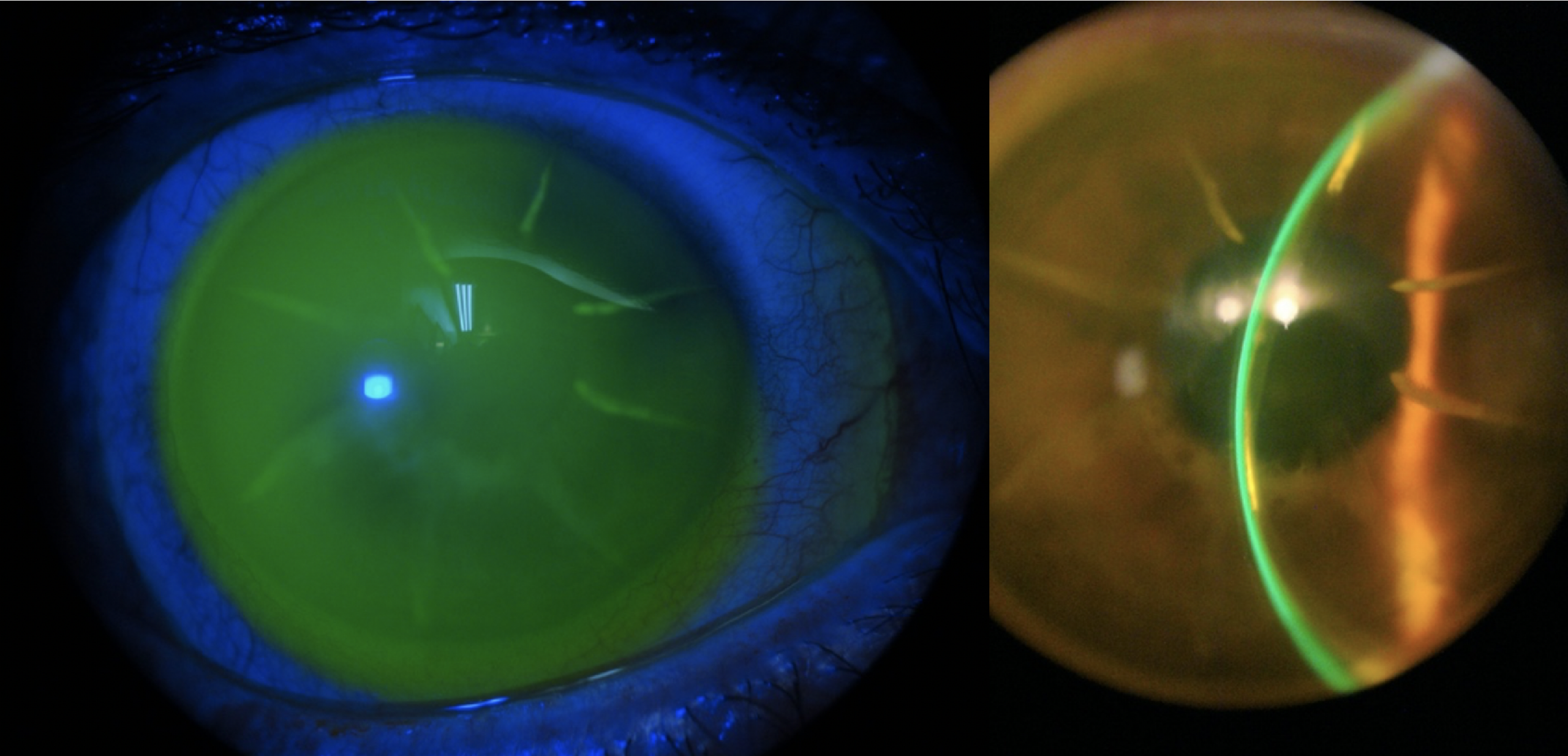
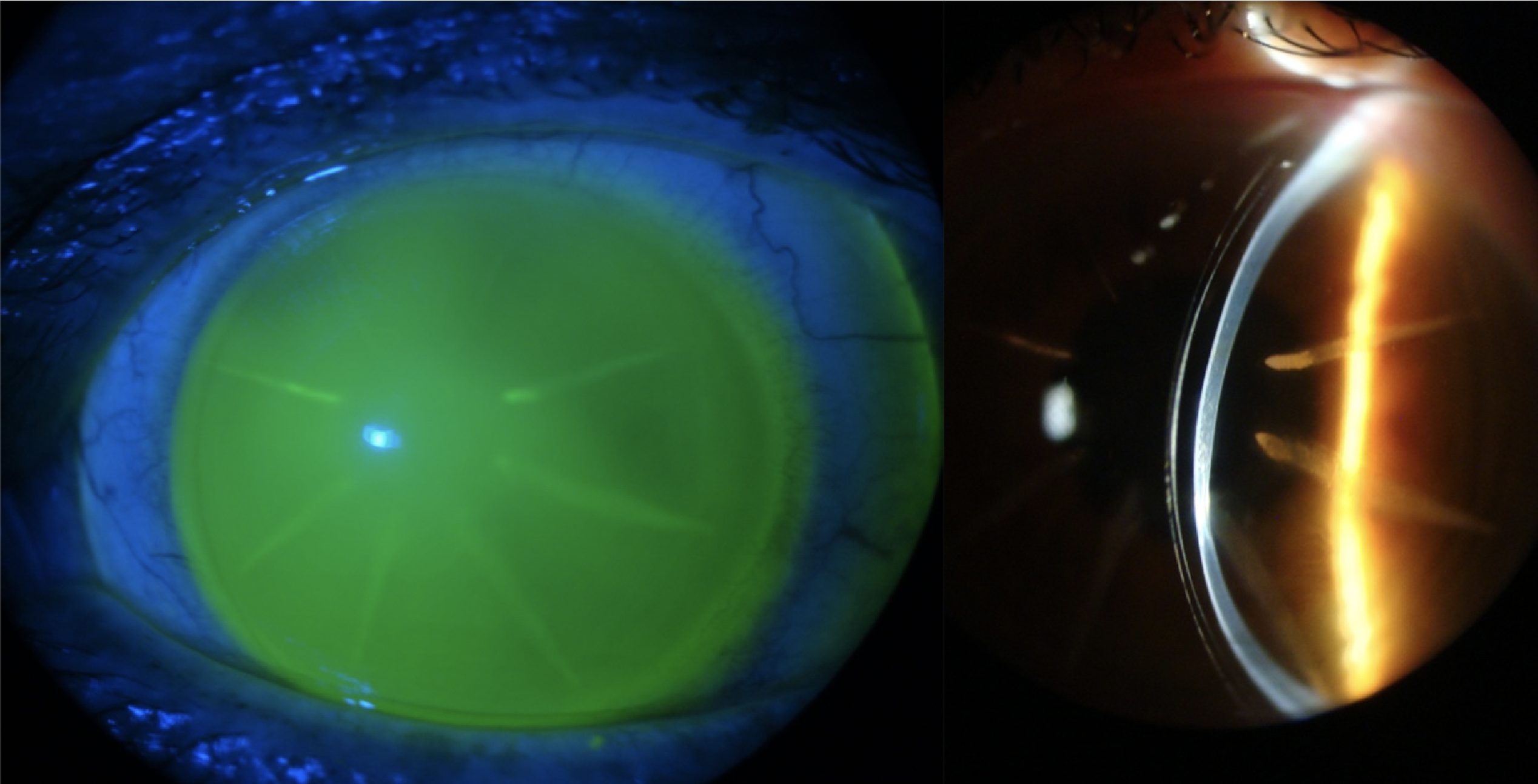
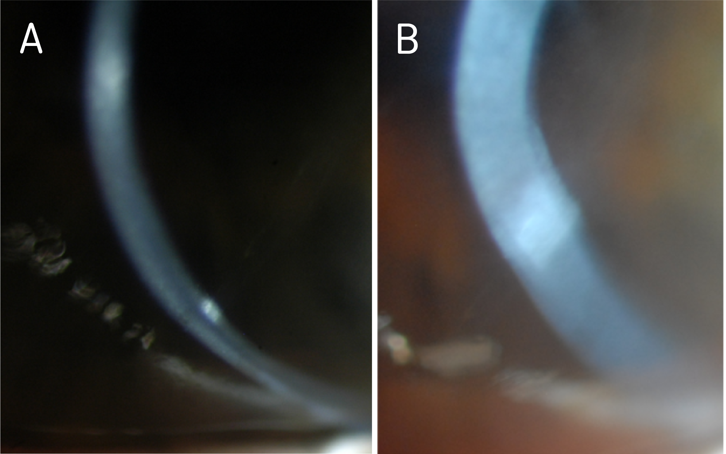
Case of a 60-year-old woman with a history of keratoconus and dry eye syndrome in both eyes, history of corneal scraping in the left eye 25 years prior, history of post hydrops left eye, and questionable potential history of amblyopia left eye. She had a history of successful PROSE treatment since 2010 (11 years of PROSE device wear) but underwent penetrating keratoplasty (PKP) in the left eye in December 2019; secondary to diffuse opacification (Figure 1) and limited best corrected visual acuity.

Prior to PKP in the left eye, she had been wearing 18.5mm, quadrant-specific PROSE devices in both eyes, with added front-surface astigmatism in the right eye and with resultant best corrected visual acuity (BCVA) of 20/30, PH 20/25-2 and 20/100, PH 20/60–, right eye and left eye respectively. With prescribed polycarbonate spectacles to correct residual astigmatism in the left eye and to serve as protection (especially for the better-seeing eye), BCVA in the left eye could be improved to 20/70. The patient wore the spectacles as needed, depending on daily activities and especially during night driving.
Reducing Suction and Maximizing Oxygen Permeability

Figure 1. Diffuse haze and opacification in the left eye, as seen on optic section (A) and wide beam (B), prior to PKP.

After corneal transplantation and having been cleared to resume scleral device wear in the left eye, she was referred to our clinic for refitting.

Evaluation After Penetrating Keratoplasty

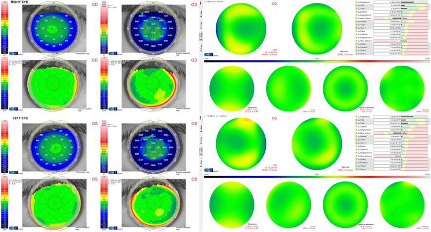
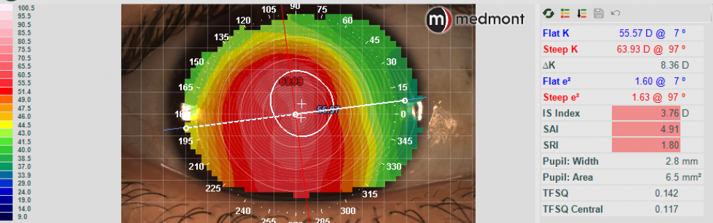
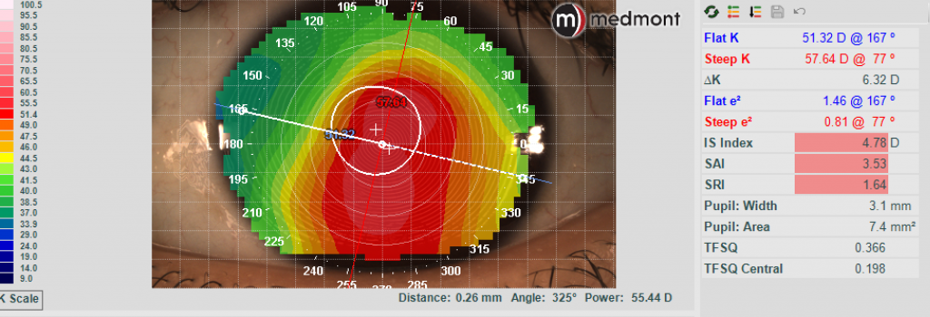
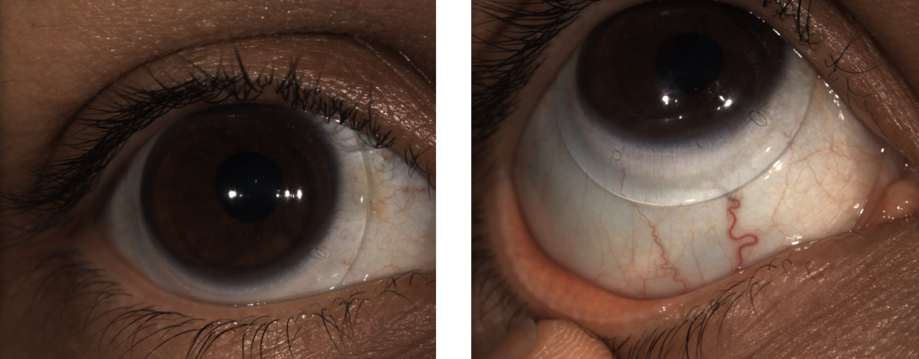
Initial evaluation after PKP OS (Topography shown in Figure 2A), showed a clear and compact graft, with interrupted sutures (Figure 2B). There was no evidence of subepithelial infiltrates, microcystic edema, no keratic precipitates, or stromal diffusion after corneal staining. The patient was on Prednisolone acetate 1% gtts QD and as sutures were removed, she was prescribed Moxifloxacin 0.5% TID in the left eye. Evaluation was also remarkable for cataracts in both eyes, not visually significant at the moment. Best corrected vision during this visit in the right eye was 20/40, PH 20/30-2 and uncorrected visual acuity in the left eye was CF at 3’, PH 20/200.
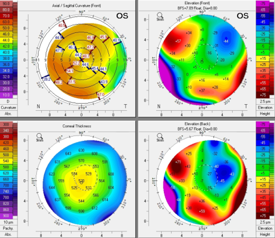
Figure 2A. Pentacam images s/p PKP in the left eye.
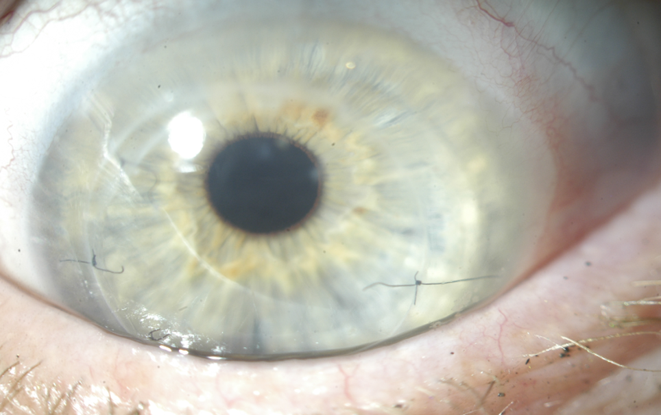
Figure 2B. Presentation of left cornea, status post corneal graft during baseline evaluation for refitting.

Refitting

We started a new fit, deciding to keep the patient in the habitual diameter of 18.5mm. Because the cornea was now grafted, we opted to design the lens using Optimum Infinite material (180 Dk) so that together with a center thickness of 0.25mm, oxygen permeability was maximized. Best documented corrected visual acuity in the left eye was 20/30-2 with the new 18.5mm device. While the patient was elated about the newfound visual potential, she reported sensation of pressure during device wear in the left eye – despite no objective evidence of significant compression at the haptics (Figure 3).
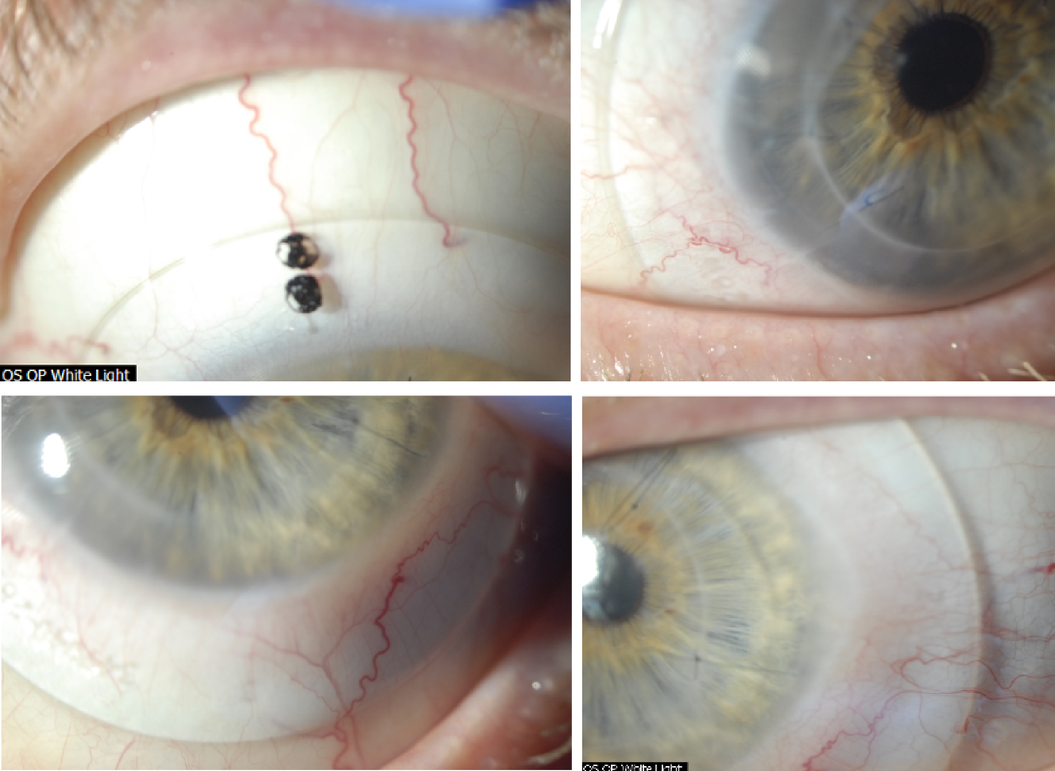
Figure 3. Initial fitting endpoint with an 18.5mm PROSE device after PKP OS.

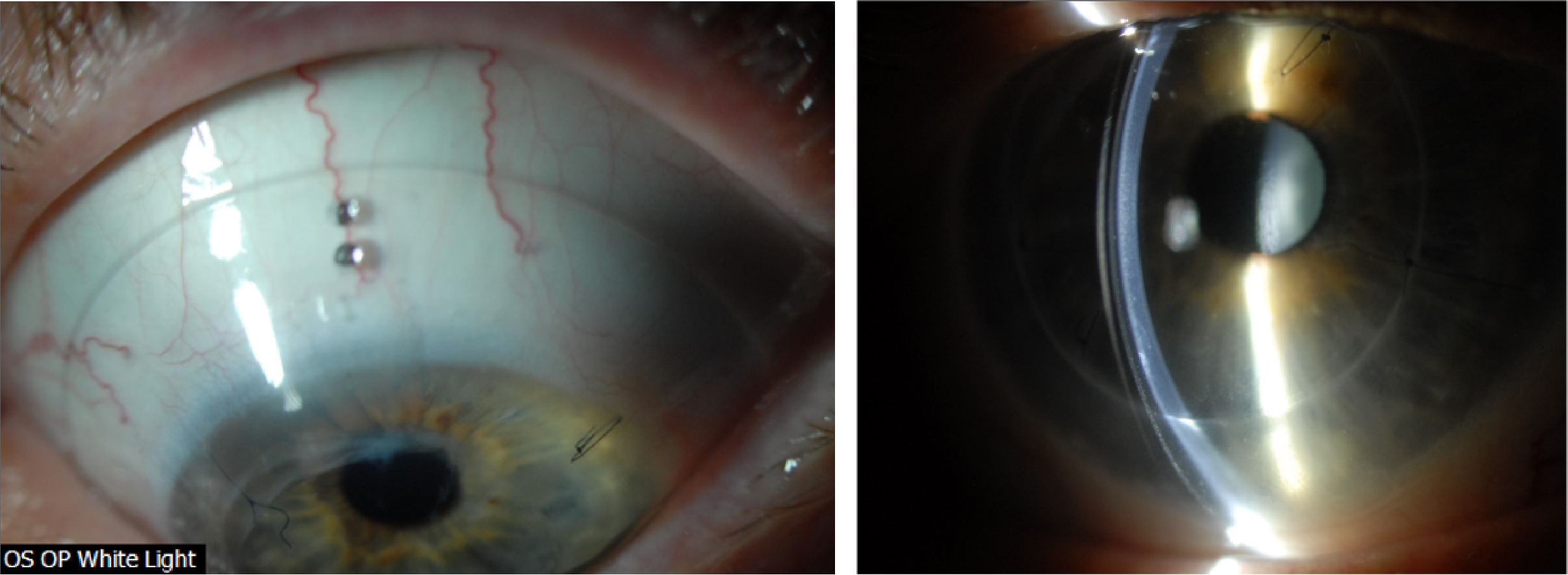
An attempt was made to flatten the haptics, modify the area along the mid-haptic portion to minimize mid-haptic compression and obtain better alignment. Despite these efforts, the patient continued to report pressure sensation with device wear and difficulty with device removal at the end of the day. These symptoms are traditionally associated with lens compression and suction with increased wear time. After reviewing the findings and the thought process with the patient, the decision was made to increase the lens diameter to 19.5mm to provide a larger surface area on which the device could rest. Figure 4 shows the new fitting endpoint with the 19.5mm, 8-meridian quadrant-specific device. One can observe good haptic alignment and the eye is white and quiet, with a nice and even distribution of lens clearance over the grafted cornea.
Reducing Suction and Maximizing Oxygen Permeability

Figure 4. Fitting endpoint with an 8-meridian, quadrant-specific 19.5mm PROSE device after PKP OS.

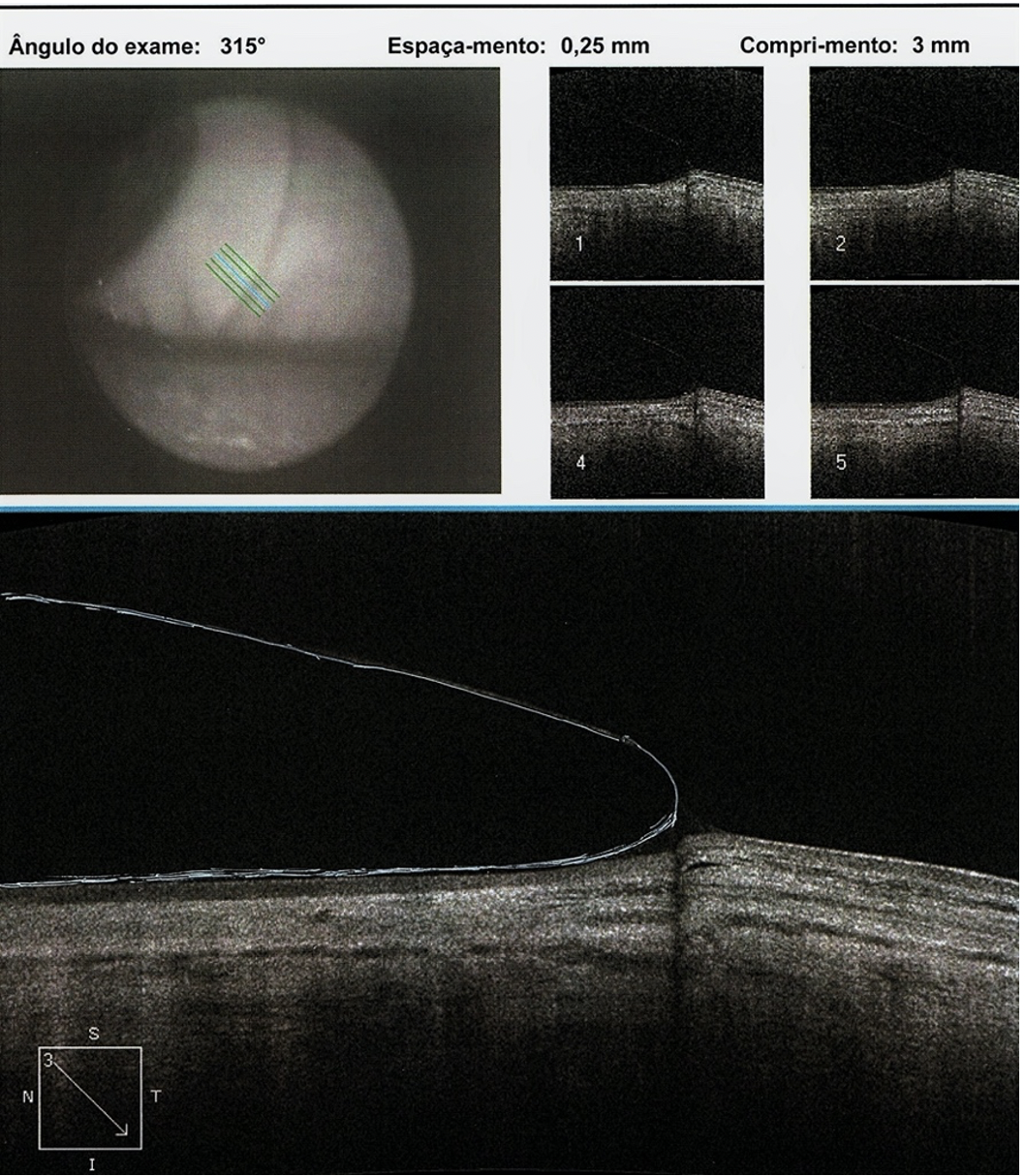
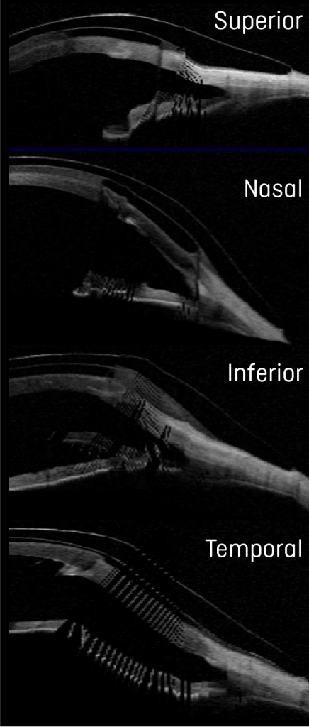
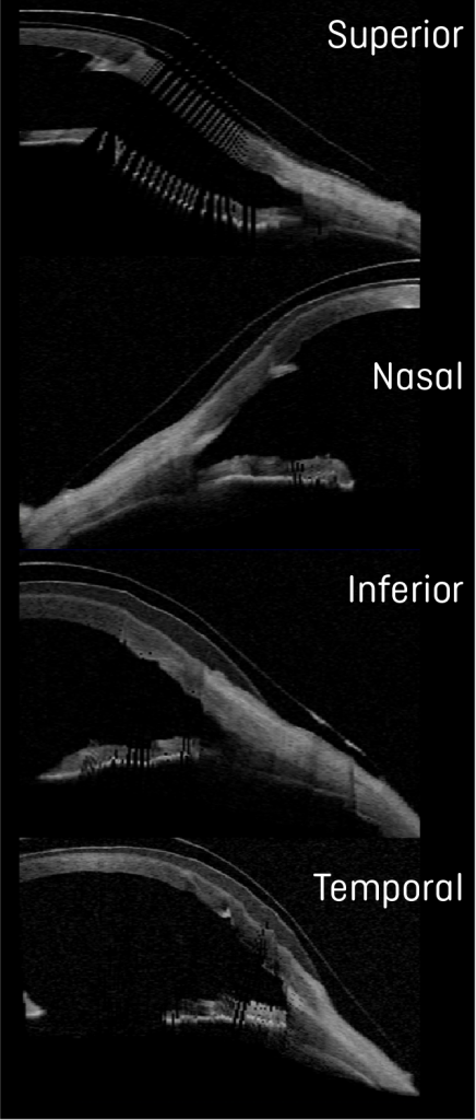
Comparison OCTs show the increased landing surface area when going from an 18.5 (Figure 5) to a 19.5mm device (Figure 6). This attempt to maximize weight distribution over a larger surface area has proven efficacious in our clinical practice when dealing with compression symptoms and device intolerance. While maintaining the same sagittal height, we additionally flattened the base curve to accommodate the more oblate nature of the cornea after transplant as shown in Figure 7.
Reducing Suction and Maximizing Oxygen Permeability

Figure 5. Ocular Coherence Tomography (OCT) of an 18.5mm device in the different orthogonal meridians of the left eye.
Reducing Suction and Maximizing Oxygen Permeability

Figure 6. Ocular Coherence Tomography (OCT) of a 19.5mm device in the different orthogonal meridians of the left eye.
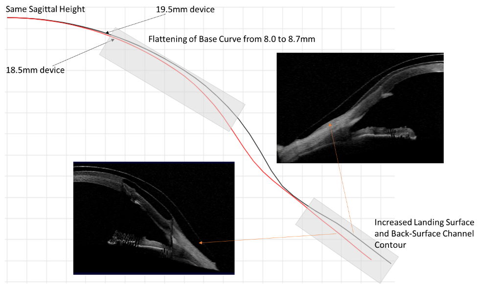
Figure 7. Graphical images of 18.5 and 19.5mm, juxtaposed to show the changes to the lens design based on parameter changes using spline curve technology and as observed by OCT. The nasal meridians corresponding to the flattest meridian in the eye are shown for comparison.

Interestingly, while we were optimizing the fit and after reporting improvement in comfort with increased lens size, the vision in the left eye dropped to the 20/50-20/60 range over the course of a couple of months. During all evaluations, we carefully monitored for signs of microcystic epithelial corneal edema, signs of rejection, etc. and at no point were evidence of these found. Given the history of previous cataracts and the fact the patient was on chronic low dose steroids, we performed an undilated assessment of the human lens in the left eye. Photo documentation revealed the development of a posterior subcapsular cataract (PSC) (Figure 8), which was later confirmed to be visually significant during dilated assessment by her corneal specialist. The patient decided to move forward with cataract extraction with hopes to improve vision.
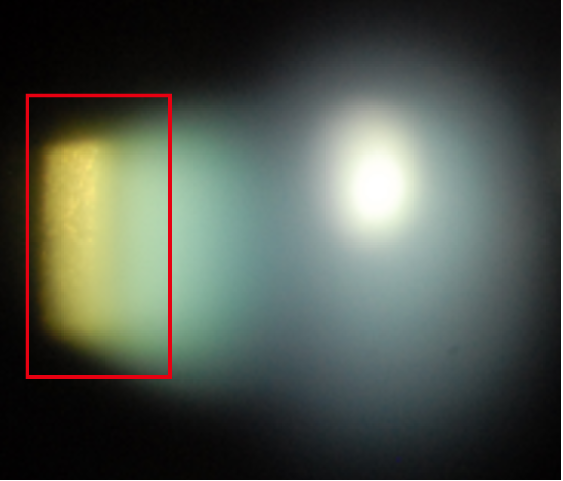
Figure 8. Evidence of PSC formation, during undilated assessment.

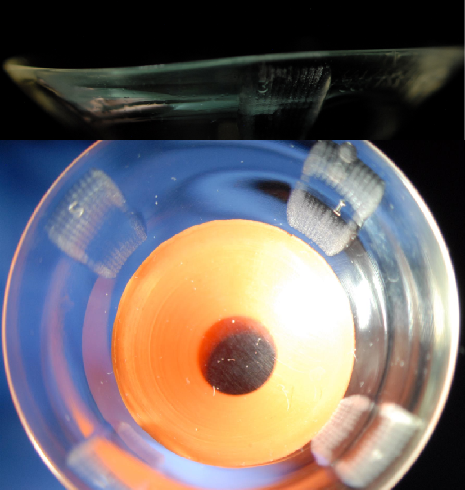
Before cataract surgery, we decided to challenge the current 19.5mm design hoping to obtain a comfortable and physiologically compatible lens design. After the patient wore the lens for one week, she reported that the previous symptoms of pressure and difficulty with lens removal had improved, but they were still present. To further minimize compression and suction symptoms, we added 4 back-surface haptic channels to soften further the landing of the haptic surface from bearing harshly on the conjunctiva and prevent a seal off effect (Figure 9).
Reducing Suction and Maximizing Oxygen Permeability

Figure 9. A 19.5mm, 8-meridian quadrant-specific PROSE device with the addition of 4 back-surface haptic channels to minimize suction and improve comfort.

Discussion and Final Outcomes

Long-term outcomes and benefits of scleral lenses after penetrating keratoplasty have been reported in the literature – while most patients benefit from improvement in visual acuity, complications with scleral lenses, i.e. microcystic corneal edema can ensue in this patient population.1 Special consideration should be taken when fitting or refitting grafted corneas with scleral lenses to minimize the risk of corneal edema, erosions and ultimate rejection. Strategies to promote fluid-ventilation like avoiding tight-fitting edges, lens seal-off and adding back-surface haptic channels can be especially important.1-5 Clinical practice has also shown that the addition of back-surface channels can have the potential to minimize lens suction over time and lead to improvement in physiological endpoints.5-7

In this case, with an increase in scleral lens landing surface area, customized haptic in an 8-meridian quadrant-specific manner and the addition of 4 back-surface haptic channels, not only are we promoting fluid-ventilation to optimize physiological endpoints, but we have a reported significant improvement in comfort. The choice to manufacture lenses in Optimum Infinite material potentiates the oxygen permeability to the grafted cornea. With the final lens after cataract surgery, the patient was able to achieve 20/25+2 best corrected visual acuity with an average comfortable daily wear of up to 12 hours.

By undertaking strategies to progressively soften the landing over the conjunctival surface to reduce or eliminate lens compression over time and minimize suction we were able to achieve a successful refitting outcome.


1. Severinksi, B. et. al. (2014) Scleral contact lenses for visual rehabilitation after penetrating keratoplasty: long-term outcomes. Cont Lens Ant Eye, 37, 196-202.
2. Rosenthal, P. Croteau, A (2005) Fluid-ventilated, gas-permeable scleral contact lens is an effective option for managing severe ocular surface disease and many corneal disorders that would otherwise require penetrating keratoplasty. Eye Cont Lens, 31(3), 130-134.
3. Severinksy, B et. al. (2010) Current applications and efficacy of scleral contact lenses – a retrospective study. J Optom, 3(3), 158-163.
4. Stason WB, Razavi M, Jacobs DS, et al. (2010) Clinical benefits of the Boston ocular surface prosthesis. Am J Ophthalmol, 149, 54–61.
5. Carrasquillo, KG, Byrnes, S (2018) Corneal edema and scleral lenses. Cont Lens Spect, 33, 34-41.
6. Barnett, M (2018) Mind the suction. Scleral Lens Monthly, Nov 2018.
7. Carrasquillo, KG (2020) Mind the suction II. Scleral Lens Monthly, Jul 2020.

Thank you to Dr Karen G.Carrasquillo for contributing to Global Insight.

[us_separator show_line=”1″ size=”small”]
[us_image image=”9526″]

Dr. Karen G. Carrasquillo is Vice President of Clinical and Professional Affairs at BostonSight. In addition, she is an adjunct clinical professor at the New England College of Optometry, an adjunct clinical professor at the school of optometry, MCPHS University, Advisory Board member for the Gas Permeable Lens Institute (GPLI), Fellow of the American Academy of Optometry (FAAO), Fellow of the Educational Society of Scleral Lenses (FSLS), and Fellow of the British Contact Lens Association (FBCLA). She is the founder and Program Chair of FitAcademy, an educational retreat for Cornea and Contact Lens Residents, and is also a planning committee member for the International Congress of Scleral Contacts (ICSC) Meeting and Global Ophthalmic Women (GLOW) meeting.
[us_separator show_line=”1″ size=”small”]
[us_post_navigation in_same_term=”1″]
[us_sharing providers=”facebook,twitter,linkedin” align=”right”]
Tags: , , , , ,

Scleral Lens Fitting Post-Refractive Keratotomy

[us_post_title][us_post_date][us_separator size=”small”]

by Luciano Bastos

Summary

Patient male, 46 years old, army official, history of post-refractive keratotomy OU performed in 1994, visual acuity was good until 2014 when it began to deteriorate. The patient had to retire early due to his condition and then began to practice law as an army lawyer. He had been wearing glasses which gave poor quality of vision and reduced acuity. He came to us from another state after visiting several doctors and testing different contact lenses in an attempt to improve his vision.

He was fitted with full scleral lenses and the results were an optimal fitting relationship, great comfort and excellent visual acuity while also preserving anterior eye and corneal physiological health.

Introduction

Our clinic has a long history of fitting irregular cornea conditions and since the early introduction of refractive radial keratotomy, many patients were referred to us in order to restore vision in cases where soft lenses or spectacles would not offer satisfactory results. During the early 90’s we relied upon the 12.7 mm wide RGP intra-limbal designs to provide the larger overall diameter to resolve cases like this. It was always a challenging process but our efforts did help many patients.

By 2014 we were able to utilise a full scleral lens design we developed after many years of research into the manufacturing and fitting of scleral lenses. My father, Dr Saul Bastos, MD asked me to study scleral lens technology and suggested that I contact his professor and friend Dr Perry Rosenthal, MD PhD (US) who became my mentor on the subject for years. I also relied on a great friend and specialist in the UK, Dr Ken Pullum FC Optom DipCLP.

During the first experimentations on manufacturing scleral lenses in 2006, we only were able to manufacture up to 16.0 mm. In the following years, new materials and diameters were available which allowed us to manufacture scleral lenses as large as 21.0 mm. There are some cases in which large, full scleral lenses are necessary, especially when there is a disease like dry eye or a large cornea with a large palpebral aperture.  

In this case, we faced an irregular corneal condition with a large cornea and palpebral aperture which clearly needed a higher SAG and a wide landing zone. The fact that it was a post-surgical cornea that had been damaged by incisions and now had scars together with reduced mechanical strength due to damage to the collagen fibres also made me aware of the importance of oxygen delivery to the cornea. At the time of the fitting, we were working with the Optimum Extreme (125 DK) from Contamac, it was reassuring to be able to rely on a higher oxygen permeability as this case required full scleral lenses.    

Examination

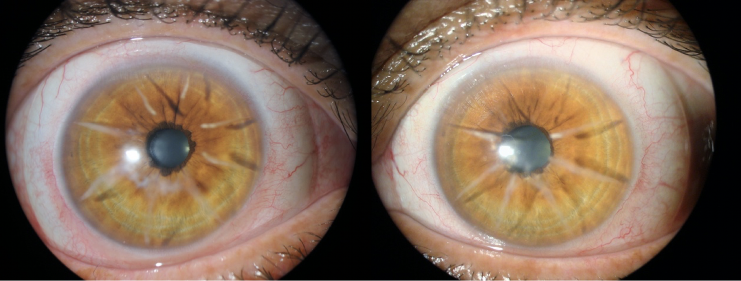
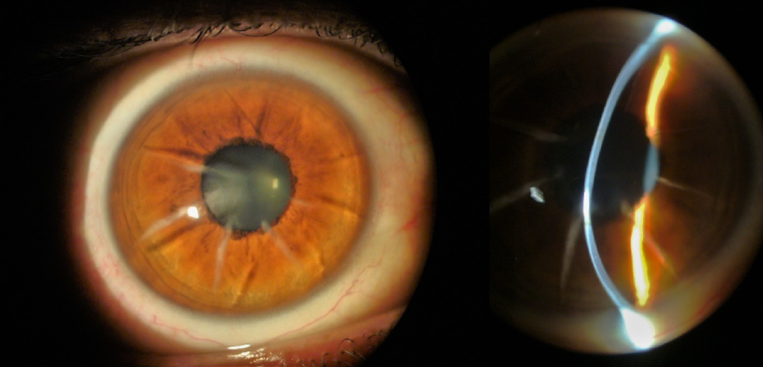
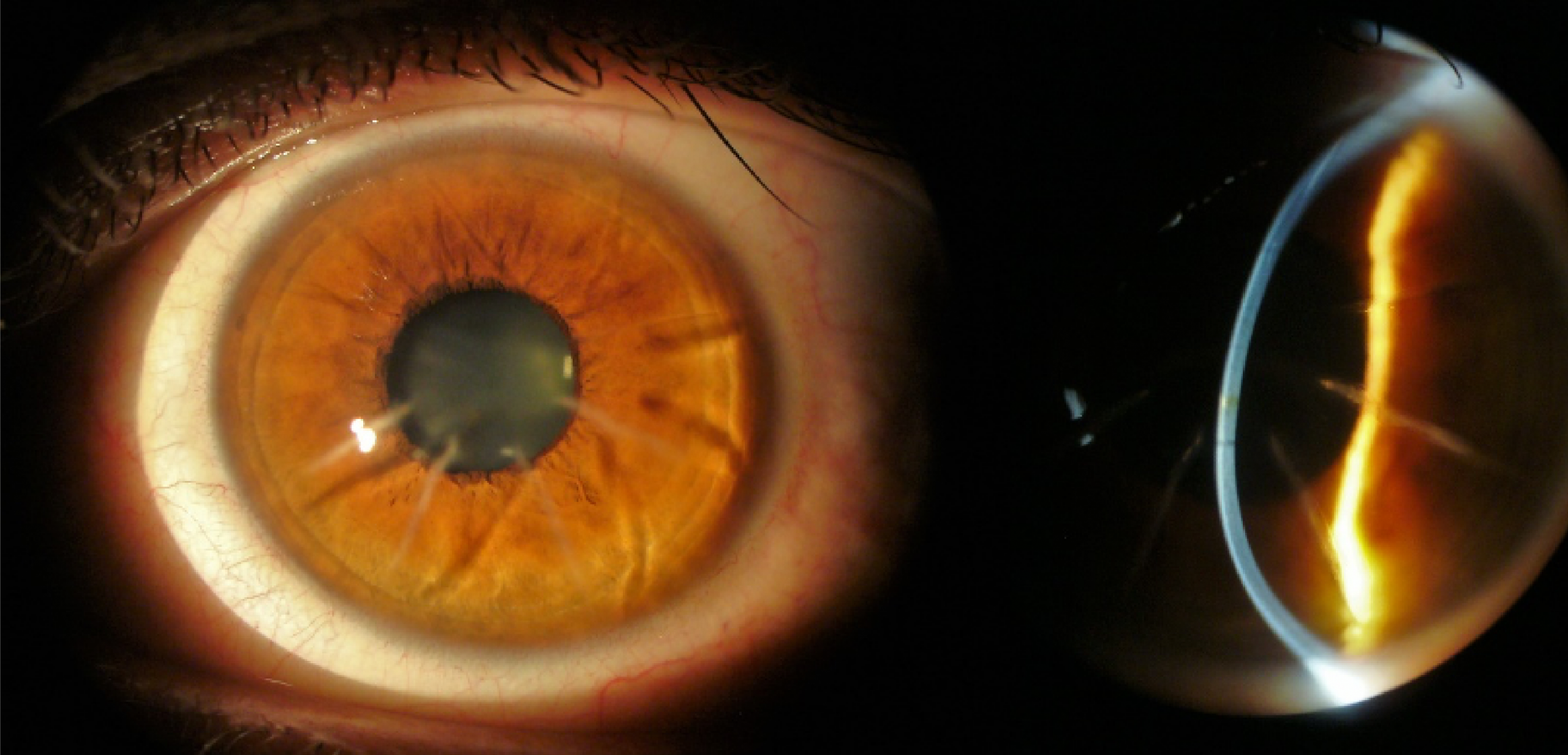
Biomicroscopy showed a series of eight radial incisions with significant depth OU and flatter topography at the central cornea. A decreased corneal thickness was also observed at the central and paracentral cornea. The slit-lamp cross-section view showed an abnormal corneal profile (Figures 1 to 4).

Scleral Lens Post-Refractive Keratotomy

Figures 1 and 2: OD radial incisions and irregular cornea profile under slit-lamp.

Figures 3 and 4: OS radial incisions and irregular cornea profile under slit-lamp.

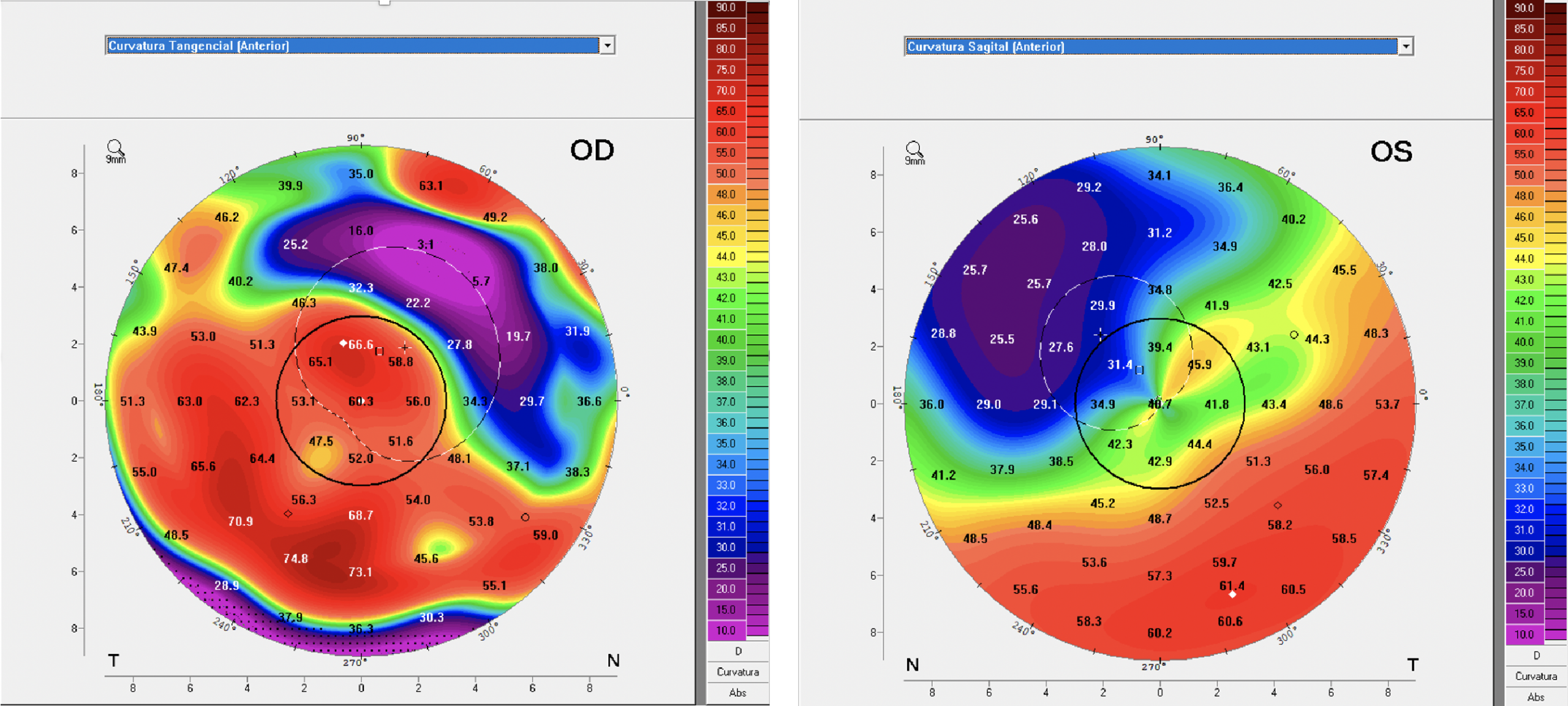
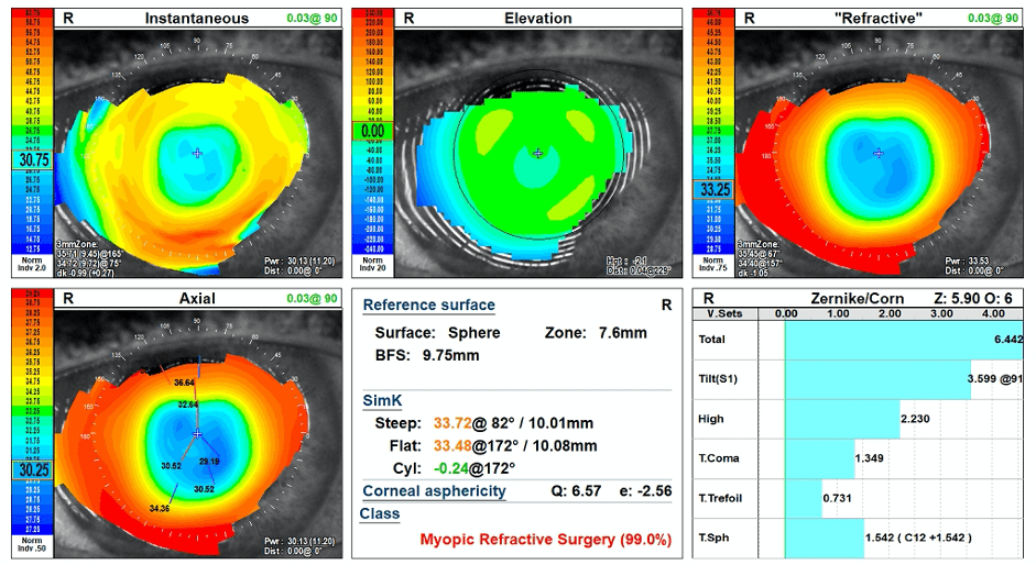
Endothelial cell count was lower than normal in both eyes, worst at OD, so we definitely would need a high Dk, a full scleral lens design and the system I developed called Wave Spline Technology (WST). WST is a ventilation channel system that allows tears to enter the fluid lens reservoir to help to deliver more oxygen to the cornea. We also performed a Pentacam Scan (Figures 5 and 6) and collected data from the precise maps this technology offers together with an OCT to verify each and every quadrant to optimise the landing haptic for a gentle and smooth fitting.    
Anterior Sagittal Curvature

Figures 5 and 6: Anterior Sagittal Curvature large maps.     

The patients that submitted to corneal refractive procedures were normally higher myopes and the larger amount of correction required necessitates deeper and more numerous incisions. Back in the day when this procedure was performed, the initial protocol was four radial incisions and, if additional correction was needed, an extra four radial incisions were made. I remember being in an ophthalmology congress with my father, Dr Saul Bastos, at which some surgeons were debating this technique and one of them said that he would perform a total of 12 or more incisions. That was really surprising to my father and me, as we were seeing those patients with those outcomes.

The procedure was abandoned with the advent of laser technology, which also has its complications. The reason I highlighted high myopia is that these patients should be carefully evaluated for glaucoma and retinal diseases as they are more prone to have posterior eye complications. Dr Marcelo Bittencourt MD (IOSB clinical director) examined the patient as a standard procedure with awareness that he was a high myopic patient before refractive surgery.      

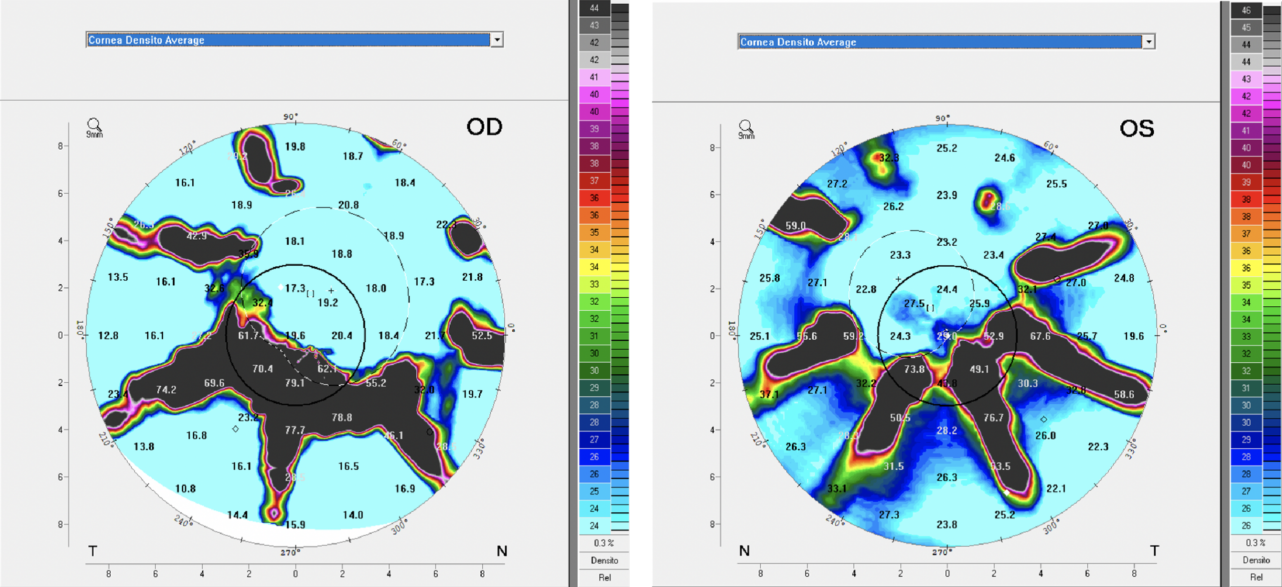
With the Density Average map from Pentacam (Figures 7 and 8), we can also observe the areas of the incisions (in black) with severe opacities. These areas also show the extensions of the incisions and its impact on the corneal epithelium and the corneal topography. The densitometer screening also helps to identify if corneal opacities or haze would change in subsequent examinations.   

Density Average maps

 Figures 7 and 8: Density Average maps identifying opaque areas at the incisions.

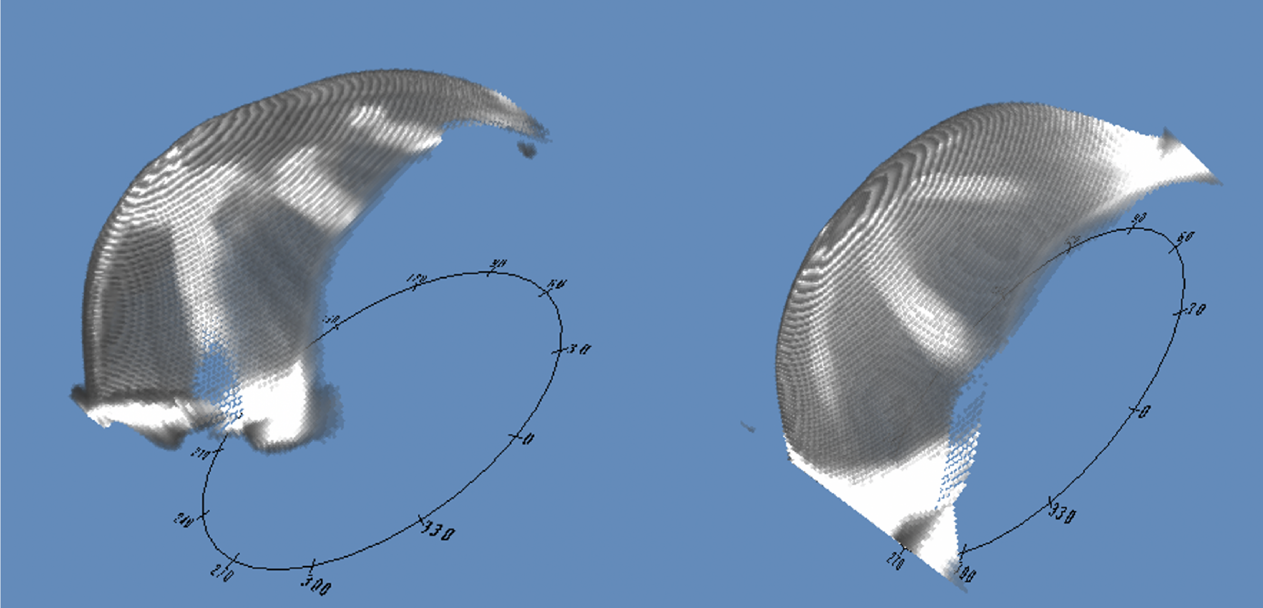
The Scheimpflug and the 3D model images are also great tools to examine abnormal cornea conditions with high irregular astigmatism. The Scheimpflug is similar to the cross-section-view on the slit lamp but it can show the cross-section in different angles. The 3D cornea model is capable of giving a better comprehension of the overall aspect of the anterior cornea and also is capable of showing areas of opacities, it can be rotated in real-time so you can view it from different perspectives, (Figures 9 and 10).

3D Corneal Model images from Pentacam

Figures 9 and 10: The 3D Corneal Model images from Pentacam, OD and OS.

We do not hesitate to use all the technology we have to get a comprehensive understanding of the case and data we can use to achieve the best possible speciality lens fit.

Contact Lens Examination and Fit Evaluation

This patient was fitted for the first time in 2014 with 19.6 mm scleral lenses. He wore the lenses for three years with no complications and came for a refit in 2017. Both fits were with the Optimum Extreme material (125 Dk).

In 2019, the patient came for a follow-up and I discussed the new Optimum Infinite material from Contamac that has a Dk of 180. The patient was happy to know more about it, and subsequently, he was refitted with the Optimum Infinite material. The lens parameters were: 

  OD OS
BC 47.50 x 45 (7.10 x 7.50) 44.00 (7.67 mm)
Power -15.00   -8.50
OAD 19.6 mm 19.6 mm
SAG  7.711 7.270
Material Optimum Infinite Optimum Infinite
Scleral Lens Post-Refractive Keratotomy

Figures 11 and 12 front images of the model SB Full Scleral lens, OD and OS.

Observe OD the enlarged opacity below the pupil, also visible in the density average map from Pentacam (Figures 7 and 11). The OS image (figure 12) also shows the visible incisions from the refractive keratotomy and it also can be compared to the Density Average image (figure 8) from Pentacam.

Initial thoughts were to design a reverse scleral lens design with a flatter posterior central base curve and a secondary, steeper base curve to overcome the flatter pattern at the central cornea. However, we were able to fit a double base curve, adjusting the base curve radius and optical zone diameters to achieve a good fit on OD as seen in figures 13 and 14.

At OS we designed a single base curve with a large optical zone diameter and we could overcome all the elevations on the cornea (Figures 15 and 16). Both eyes were fitted with a SAG that proved to be high enough after settling with four hours of lens wear. The frontal and cross-section view observed and the slit lamp shows the fluid lens reservoir at both perspectives.     

Scleral Lens Post-Refractive Keratotomy

Figures 13 and 14: OD frontal and cross-section view on the slit lamp with fluorescein.

Scleral Lens Post-Refractive Keratotomy

Figures 15 and 16: OS frontal view with fluorescein and cross-section view with white beam.

The patient was happy with the comfort, and visual acuity was 20/10 J1 OU. The visual outcome was much better than expected despite the fact he is 46 years old, (so he should be entering presbyopia soon) he was able to read J1 with no add correction. He was informed about the possible future need of add correction, we determined his dominant eye for possible near vision correction when necessary, utilizing reading glasses, monovision or a multifocal design in the early stages.          

We also performed an OCT scan at eight different angles to have a better understanding of the landing pattern in each meridian. The scans were separated by each angle; one image of the limbal transition and the other of the landing haptic including the edge as you can see in figure 17.

The OCT exam is a great tool to identify areas where the haptic may present significant pressure over the conjunctival sclera, or excessive lift. With all information gathered from these 16 images generated by OCT we can plan and design the scleral lens with an estimation of toricity that we may apply to the haptic and align where required.

Scleral Lens Post-Refractive Keratotomy

Figure 17: A single segment of the haptic showing scleral alignment.

Follow-up Visit After 120 Days

During the weeks following the dispense, the patient reported feeling good, with excellent vision. After about 120 days of dispensing, he was able to visit us again, bearing in mind he lives in another state.

Biomicroscopy during examination showed a crystal-clear cornea, specular biomicroscopy remained stable with a similar endothelial cell count. Visual acuity also remained stable and there was no sign of corneal edema or any complications. Our normal procedure during follow-up visits is to re-evaluate the fitting and consider what might be required to improve the overall fitting. In this case, we decided that with the next lenses we would increase the landing zone and also slightly augment the transitional limbal zone from the base curve to the haptic landing zone. This will allow the lens to have a better pattern over the limbus as it appears to be too close to the limbus (figures 13 and 15).
Customising the Scleral Lens Fitting

When we change any variable at the posterior curve, the optical zones and the overall diameter of the scleral lens SAG are affected. The more variables you change the more impact it has on SAG value, achieving a lower or a higher value depending on the changes made. If you have, like in this case, a SAG that you have as a reference it is a great starting point as you will need to apply the changes to customize the fitting. So, it is necessary to control these variables in a manner that the changes will result in a better fitting relationship while preserving the SAG value to a number close to the reference.     
This is absolutely true with trial lenses as well; we always think about customizing the fit whenever it is necessary. If the practitioner does not have software to calculate these changes it is of utmost importance to have technical assistance from a lab consultant that may be able to provide the possible calculations to achieve the results the specialist wants. Another possibility is to use what Ultralentes call Digital Consultancy. In this case, the doctors are encouraged to produce frontal and cross-section view images through the slit-lamp and forward these photos or short videos to the lab. This is often the case as practitioners generally do not use contact lens software to design lenses. Ultralentes also provide the practitioners with a Consultancy Form that the doctor may fill and also make observations and drawings of the specific areas he wants to improve from the trial lens used. 

Conclusion

The patient reported feeling really great with his new lenses, he mentioned that he was happy to change to a higher Dk material (Optimum Infinite). He reported greater comfort, visual acuity remained stable and the eye calm. The overall fitting was quite acceptable for a surgical cornea that has multiple incisions that had deteriorated the corneal physiology over the years.             

We know that patient’s previous lenses were made in Optimum Extreme and were worn for two years, they were fine and the motive for a new fit was based on taking advantage of higher oxygen transmissibility rather than necessity. Although the lenses were still acceptable to wear, naturally, a new pair may impact visual quality as acuity had no change. What is important is that after four months of wearing the new material, the results were clinically excellent and the patient was happy with comfort and vision.             

I also want to address that we had no problem manufacturing these full scleral lens designs in the Optimum Infinite material. It proved to be stable with excellent physical properties with a 180 Dk. This is really important especially when these speciality lens designs are planned.

At the time of writing this case, we already have several scleral lens fittings with Optimum Infinite and it is definitely a great and reliable material from Contamac. The higher Dk material for scleral lens patients means an extended wearing time but is especially important to post-surgery patients.

Here at the Instituto de Olhos Dr Saul Bastos (IOSB), 96% of the fittings are speciality RGP and scleral lens fittings. From these, about 70% are ectasic patients, including (most) keratoconus patients, pellucid Marginal Degeneration and keratoglobus. About 15% are post-surgical patients like post-penetrating keratoplasty (corneal transplants), post-radial keratotomy and post-laser surgery (Lasik and PRK), about 5% are dry eye patients. The remaining 10% of patients are moderate to high myopics, hyperopics, and aphakics. The introduction of the Optimum Infinite material is a great advantage to these post-surgery patients and those with the larger full scleral lens designs.  


1. Banta JT. Ocular Trauma – Bascon Plamer Eye Institute. Miami, FL – Saunders Elsevier, 2007
2. Black CJ, Rosenthal P, Kaufman HE et al. Symposium on Contact Lenses. Transactions – New Orleans Academy of Opthalmology. St Louis: Mosby, 1973
3. Bowden TJ. Contact Lens: The Story – Cornwall, UK. London: Bower House, 2009
4. Cochet P, Amiard H. Contact Lens International Clinics. Boston: Little, Brown, 1969
5. Cvintal T, Munarin MAM, Moro F. Complicações do Transplante de Córnea. São Paulo, Santos, 2004
6. Duker JS, Macsal MS, Fontes BM. Anterior Segment – Rapid Diagnosis in Ophthalmology, St. Louis, Mosby Elsevier, 2008
7. Girard LJ, Soper JW, Gunn C. Corneal and Scleral Contact Lenses. Proceedings of the International Congress. St. Louis: Mosby, 1967
8. Girard LJ, Soper JW, Sampson WG. Corneal Contact Lenses. St. Louis: Mosby Co. 1970
9. Godinho C, Dantas B, Sobrinho M.et al. O Padrão CG em Lentes de Contato. Bastos, L. Cultura Médica;. pág. 301 a 313. 2010
10. Jenkin L, Tyler-Jones R. Theory and Practice of Contact Lens Fitting: Hatton Press 1969
11. Pena A. Bastos, S et all, Clínica de Lentes de Contato. Cultura Médica, 1989; pág. 69-79
12. Philips AJ, Speedwell L, Morris J. Contact Lenses – 5th ed. Ed Butterworth Heinmann Elsevier 2007
13. Ferrara, Paulo.  Anel de Ferrara – Cultura Médica – 2006
14. Romero-Rangel T, Stavrou P, Cotter J et all. Gas Permeable Ocular Prosthesis Therapy in Ocular Surface Disease. Am J Ophthalmology, 2000, 130: 25-32
15. Rosenthal P, Cotter JM, Treatment of Persistent Epithelial Defect with extended wear of a fluid-ventilated gas permeable scleral contact lens Am J, Opthalm 2000; 130: 25-32
16. Rosenthal P., Cotter JM. The Boston Ocular Surface Prosthesis in the management of Severe Ocular Surface Disease. In: Foulks G. (ed.) Ophthalm Clin A Am 2003; 16: 89-93
17. Schein OD, Rosenthal P, Ducharme C. A Gas Permeable Scleral Lens for visual rehabilitation. Am J Ophthalmol, 1990; 109:318        
18. Fadel, Daddi DOptom, FSLS, FBCLA, FAAO – Scleral Lens Issues and Complications: Their Recognition, Etiology and Management. – Dougmar Publishing Group Inc. August 2020

Thank you to Dr Luciano Bastos for contributing to Global Insight.


[us_separator size=”small” show_line=”1″]
[us_image image=”9427″]
[us_separator]

Mr. Bastos is the director and clinical instructor of specialty contact lenses at the Instituto de Olhos Dr. Saul Bastos (IOSB), and is the director and specialty lens consultant of Ultralentes, a small laboratory specializing in GP and scleral lens designs in Porto Alegre, RS, Brazil.

[us_separator size=”large”]
[us_separator show_line=”1″ size=”small”]
[us_post_navigation in_same_term=”1″]
[us_sharing providers=”facebook,twitter,linkedin” align=”right”]
Tags: , ,

Maximising Oxygen in a Post PKP Patient

[us_post_title][us_post_date][us_separator size=”small”]

by Steven Sorkin, OD, FSLS

Patient History

A new patient was referred for specialty contact lens evaluation by his corneal surgeon.

The patient is a 61-year-old male who has a complex ocular past history. He has a history of ocular trauma to the right eye approximately 5 years prior. A wood-based foreign body had caused a corneal ulcer and subsequent dense corneal scarring which necessitated a full-thickness penetrating keratoplasty 6 months after the original injury. One year later he required cataract surgery in that eye. He subsequently developed a full-thickness macular hole in the previously normal left eye leaving him with 20/400 best corrected acuity in the left eye. Being monocular at this point, corneal GP lenses and hybrids were tried with poor success. He was referred to me for scleral lens evaluation.

Maximising Oxygen Post PKP

Evaluation

Entering uncorrected visual acuities were counting fingers in both eyes.

Refraction in the right eye was -6.00 +7.00 x 075 20/50. The left eye did not improve past 20/400.

Baseline pachymetry was 650 microns in the right eye.

Maximising Oxygen Post PKP

Specular microscopy was 1113 cells/mm squared with good CV and HEX values.

K’s were 42.50/52.12 M 070 in the right eye.

Goldmann tonometry was 14/15.

Anterior segment evaluation of the right eye showed a healthy full-thickness PKP. No microcystic edema, corneal staining or keratic precipitates were observed.

Retinal exam was unremarkable in the right eye with no maculopathy.

Maximising Oxygen Post PKP

Scleral Lens Fit

A detailed discussion with the patient regarding risks and benefits of contact lens wear occurred at the consult visit. After this discussion, we commenced with a diagnostic scleral lens fit. The diagnostic lens had 296 microns of clearance after 30 mins of settling. Over refraction improved his visual acuity to 20/25-.

A lens was ordered for the patient in the following parameters:

Blanchard One Fit Med

OD central vault 5050/ Rx -5.00/ M-50/L STD/ EDGE +150/+75/Diameter 15.6/ CT 0.24

I ordered the lens in Optimum Infinite material to maximise the oxygen permeability in this post graft patient.

At the dispense visit, central vault was 192 microns and overall lens fit was acceptable. Edge configuration was good without edge lift or blanching. Visual acuity was 20/25- and the patient was very pleased with comfort as well.

Follow Up Visit

At the one week follow up visit, the patient was very happy with the lens. The fitting evaluation was excellent with a final central vault of 150 microns after 6 hours of wearing time. The mid-peripheral fitting relationship was also good with no touch. No distress to the corneal transplant was noted. Central corneal thickness was noted at 665 microns, a nominal increase from baseline. No further lens adjustments were needed. The patient can wear his lens greater than 12 hours per day. A six-month evaluation also showed no change.

We are following him every three months to monitor his corneal transplant and perform sequential pachymetry, specular microscopy and topographies to watch for any adverse effects or complications going forward. For this patient, utilising a hyper Dk lens material and minimising lens thickness, along with a proper fitting lens improved the quality of vision in this monocular patient that has a challenging ocular history.

Thank you to Steven Sorkin, OD, FSLS for contributing to Global Insight.

[us_separator size=”small” show_line=”1″]
[us_image image=”9433″]

Dr. Sorkin serves as the Director of Specialty Contact Lens Services at Corneal Associates of New Jersey in Fairfield, New Jersey. He earned his Doctor of Optometry degree from the State University of New York College of Optometry. He lectures extensively throughout the United States and internationally on contact lenses, dry eyes, ocular therapeutics and corneal diseases. He has a special interest in difficult to fit contact lens patients, such as those with keratoconus and post surgical cases and manages many patients with complex anterior segment conditions. He participates in many contact lens related studies. Dr. Sorkin currently serves as President of the Essex County Optometric Society and is a  member of the Board of Directors of the New Jersey Society of Optometric Physicians.  He is also a Fellow of the Scleral Lens Education Society and is adjunct faculty at Salus University. He was named New Jersey Optometrist of the Year by NJSOP in 2018.

[us_separator size=”small” show_line=”1″]
[us_post_navigation in_same_term=”1″]
[us_sharing providers=”facebook,twitter,linkedin” align=”right”]
Tags: , ,

Successful Visual Restoration of Presbyopia with a New Customized Multifocal Corneo-Scleral Contact Lens

[us_post_title][us_post_date]

by David P Piñero, PhD

Introduction

The use of scleral contact lenses has grown significantly in the last year, with an exponential increase in research on this issue.1 Although there is a consensus about most of the aspects of contact lens management among practitioners with more than 5 years of scleral lens experience,2 there are some aspects that require more research, such as the management of multifocality for presbyopia correction. Recently, a new model of multifocal corneo-scleral contact lens has been commercially released by Laboratorios Lenticon S.A. (Madrid, Spain), Presbycustom, providing the option of customizing not only the fitting parameters to adjust the lens over the cornea and sclera, but also the type of multifocality induced. We report the results obtained with this new modality of multifocal scleral contact lens in a specific case not tolerating multifocal soft contact lenses due to poor and fluctuating visual quality.

Case report

A 45-year old man attended the Contact Lens Unit of the Department of Ophthalmology of the Vithas Medimar International Hospital (Alicante, Spain) asking for a contact lens solution for his presbyopia. He was a contact lens user for 16 years. He noticed some difficulties at near due to the development of presbyopia and tried to find a contact lens solution for the compensation of this condition. However, the results obtained with different models of soft multifocal contact lenses (5 contact lens models were previously tried) have been unsatisfactory as the visual quality with them was not good, with fluctuation of vision with most of them. On the first examination, the following clinical data were obtained:

  Right Eye Left Eye
Subjective Refraction +1.75 -0.50 x 70 +1.00
Corrected Distance Visual Acuity (CDVA) 0.00 logMAR 0.00 logMAR
Near Addition +1.00 +1.00
Corrected Near Visual Acuity (CNBA) 0.00 logMAR 0.00 logMAR
Biomicroscopy No alterations detected No alterations detected
Scotopic Pupil Size 6.0 mm 6.0 mm
Mean Keratometry 41.17 D 41.2 D
Ocular Spherical Aberration (6 mm pupil) 0.26 µm 0.21 µm

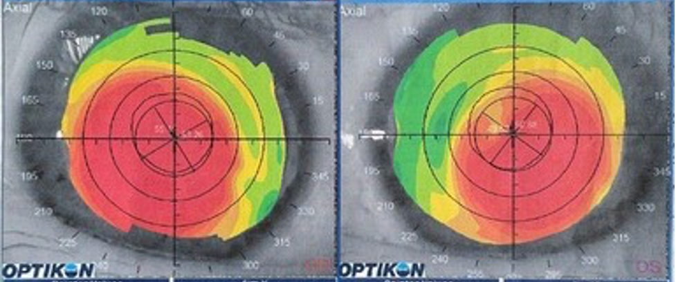
No significant corneal shape alterations were found on corneal topography, with values of corneal asphericity and high order aberrations within a normal range (Figure 1). Considering the peculiarity of the case, we decided to fit a customized multifocal scleral contact lens. Specifically, the Presbycustom contact lens was fitted, which is a corneo-scleral contact lens made of highly gas permeable material (Roflufocon D, Optimum Extra, 100 Dk). This lens has a customized design according to the anterior corneal geometry, the level of ocular high order aberrations and the peculiarities of pupil dynamics. The corneal area has a variable diameter with a fixed back surface asphericity and a central anterior surface asphericity which is modified according to the aberrometric induction required. Specifically, the increase in the depth of focus achieved with the contact lens is generated by customizing the induction of primary and secondary spherical aberration.3,4

Multifocal Corneo-Scleral Lens

Figure 1. Corneal topographic analysis with the Sirius system (CSO, Firenze) in both eyes of the case report described.

Different contact lenses were tried until obtaining the best fit with the following lenses in right and left eyes, respectively:

  Right Eye Left Eye
Lens Diameter 13.5 mm 13.5 mm
Corneal Radius 7.65 mm 7.46 mm
Scleral Radius 7.55 mm 7.40 mm
Optical Power -1.00 D -3.00 D
Anterior Eccentricity 0.63 0.55
Posterior Eccentricity 0.63 0.63

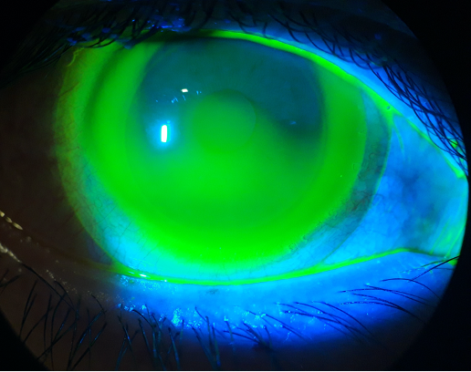
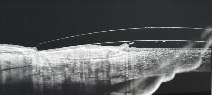
With these lenses, the fluorogram was correct, showing a central tear pooling, with a paracentral alignment and a significant edge clearance (Figure 2). Likewise, a smooth peripheral bearing of the lens was observed by means of optical coherence tomography (Figure 3).

Multifocal Corneo-Scleral Lens

Figure 2. Fluorogram obtained in the right eye of the case fitted.

Multifocal Corneo-Scleral Lens

Figure 3. Peripheral bearing of the lens observed by means of optical coherence tomography.

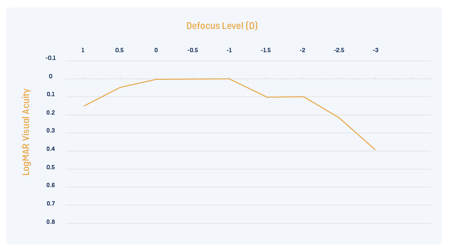
At 1 month of contact lens wear, the visual acuities with the contact lens were 0.00 logMAR and 0.10 logMAR at distance and near (40 cm), respectively, in both eyes (overrefraction: plano in both eyes). Figure 4 shows the binocular defocus curve measured, confirming the significantly enlarged depth of focus achieved.

Binocular defocus curve

Figure 4. Binocular defocus curve measured in the patient.

The patient has been successfully wearing the contact lens for 2 years, with maintenance of the results and good levels of tolerance.

Conclusion

The use of customized scleral contact lenses allows the practitioner to optimize the presbyopia correction in any type of presbyopic patient. The selection of an appropriate combination of primary and secondary spherical aberration of opposite signs according to the aberrometric status of the eye is an effective tool to optimize the depth of focus required for presbyopia correction in each individual case. This preliminary result should be corroborated in the long term as well as in a larger sample size.


1. Davies I. Scleral publications & contact lens category growth. Cont Lens Anterior Eye 2019; 42: 234-235.
2. Van der Worp E, Bornman D, Ferreira DL, Faria-Ribeiro M, Garcia-Porta N, González-Meijome JM. Modern scleral contact lenses: A review. Cont Lens Anterior Eye 2014; 37: 240-50.
3. Yi F, Iskander R, Collins M. Depth of focus and visual acuity with primary and secondary spherical aberration. Vis Res 2011; 51: 1648-1658.
4. Benard Y, López-Gil N, Legras R. Optimizing the subjective depth-of-focus with combinations of fourth- and sixth-order spherical aberration. Vis Res 2011; 51: 2471-2477.

Thank you to David P Piñero, PhD for contributing to Global Insight.

[us_separator show_line=”1″ size=”small”]
[us_separator][us_image image=”9441″]

David P Piñero has a degree in Optics and Optometry from the University of Alicante (1998) and a degree in Information and Documentation from the Open University of Catalonia (2006). He obtained his PhD Degree at the University of Alicante in 2010 with the doctoral thesis entitled “Characterization and modelling of the effect of the intrastromal ring segments de in ectatic corneas”. Likewise, he obtained the qualification of University Specialist in Pre- and Post-surgical Optometry by the University of Valladolid in 2001. Currently, he is professor/researcher at the Department of Optics, Pharmacology and Anatomy of the University of Alicante, associate editor of Journal of Optometry, Journal of Ophthalmology and BMC Ophthalmology, and optometrist at the Department of Ophthalmology of the Vithas Medimar International Hospital (Alicante, Spain). He has published more than 300 papers in peer-reviewed journals, most of them on the cornea, and participated in 20 book chapters. He has also participated in more than 20 research projects.

[us_separator show_line=”1″ size=”small”]
[us_post_navigation in_same_term=”1″]
[us_sharing providers=”facebook,twitter,linkedin” align=”right”]
Tags: , ,

Backtracking From Scleral Lenses: When Custom Soft Lenses Get the Job Done

[us_post_title][us_post_date]

by Priya Patel, OD.

Introduction

Scleral lenses provide significant visual and therapeutic advantages for patients suffering from a variety of corneal conditions: inclusive of kerato-ectasias, penetrating keratoplasties, and varying severities of corneal irregularities.

This advancement in the specialty lens realm has expanded to the use of sclerals, yet previous technologies are still capable of providing comparable comfort and vision. 

Custom soft lenses have potential to significantly improve vision without the need for complex designs, and small diameter gas permeable lenses can achieve similar visual outcomes as sclerals at a fraction of the cost. Custom soft lenses can be entirely customizable from base curve, diameter, power, to even center thickness to improve upon masking corneal irregularities. Often in clinical practice, there exist simpler solutions to complex problems.

Case Report

A 46-year-old White male teacher with a history of keratoconus OD>OS presents to our office seeking scleral lenses. Patient JB has a history of gas permeable (GP) lens intolerance and has successfully avoided PKP (penetrating keratoplasties) in both eyes. The continuous irritation in his GP lenses and the fear they can pop out at any moment, has him seeking out scleral lenses.

His corneal shape shows most significant steepening directly over the visual axis, which would be well vaulted by a prolate design. Utilizing the Zenlens (Alden) Prolate fitting set, JB was fit into a 16.0 diameter scleral lens in both eyes, achieving BCVA 20/25 OD/OS. The initial fitting period was completed without any significant complications, and JB was happy to have clear, comfortable vision. At the one year mark after his initial scleral lens fitting, JB started to develop significant redness after 4 hours of lens wear with concurrent fogging, which only resolved after reinsertion of his lenses. After lens removal, the chronic irritation persisted. This led to intolerance of his lenses.

Zenlens Prolate
Scleral Lens
Right Eye Left Eye
SAG/Base Curve/Diameter (μ/mm/mm) 4950/6.70/16.0 4950/6.70/16.0
Power -4.75-2.00×150 -6.25-1.25×070
Limbal Clearance Curve (LCC) +60 STD
Advanced Peripheral System H = 7 steps flat
V = STD
2 steps flat
Visual Acuity 20/25 20/25

JB became more frustrated, and he pleaded for any lens option, which would yield a similar visual outcome, but improve his comfort. We were sceptical we would be able to achieve this, but in an effort to exhaust all options available,  we switched him into a custom soft lens modality in both eyes.  To optimize comfort and reduce the chance of redness, fogging, irritation after lens removal as JB previously experienced, he is fit into the NovaKone Toric OD and Biofinity XR Toric OS. The custom soft lens selection was based on the poor BCVA (20/80+) attainable through refraction: the customizable center thickness of the Novakone lens would allow greater control in neutralizing irregular astigmatism in the right eye. The left eye was correctable to an adequate level (20/25+) with glasses so a monthly disposable lens had potential to correct JB to a level at which he would be functional. 

Custom Soft Lenses

K Values:

OD: 55.15@121/53.26@031

OS: 50.88@053/49.52@143

Custom Soft Lenses

OD: Novakone Toric lens, rotated 15 degrees temporally (markings are highlighted with yellow dashes).
OS: Biofinity XR Toric lens, showing no rotation (lens marking is highlighted with a yellow dash).

 
Refraction  
OD -12.75-1.75×025      20/80+
OS -0.50-4.00×145       20/25+
Custom Soft Lens Right Eye Left Eye
Name Novakone Toric Biofinity Toric XR
Base Curve/
Diameter (mm)
7.8/8.4 IT=2 8.7/14.5
Power -7.00-0.75×020 -2.00-2.75×160
Visual Acuity 20/20- 20/20-2

These changes in lens modality improved his contact lens wearing experience and significantly simplified his daily lens wear routine. He has been in his new lenses for over a year with no degeneration in vision, and no recurrences of discomfort or irritation.

Discussion

Scleral lenses are a mainstay as a rehabilitative and therapeutic contact lens option, but alternatives exist for patients from mild to advanced cases of corneal irregularities. Taking into account comfort, lens handling capabilities, alternative specialty lens considerations can lead to improved outcomes as demonstrated in this case.

Scleral lenses require hand steadiness, ability for full control of fine motor skills, and neck mobility. Patients suffering from tremors, neck pain, or mobility issues often have trouble inserting scleral lenses on their own, but have minimal issues with soft and GP lenses. Improper insertion of scleral lenses can lead to air bubbles, and discomfort with lens wear. On the contrary,  minimal risk is associated with difficulties with soft lens insertion .¹

Scleral lenses are associated with an increased risk of hypoxia if the tear film layer between the corneal surface and the back surface of the lens exceeds 200nm.²?³ Soft and GP lens lenses pose a lower risk for creating a hypoxic environment even for fragile corneas.

Utilizing higher modulus soft lenses allow some masking of irregular astigmatism for better visual outcomes. Although in this case, since JB achieved adequate vision through a refraction  in his left eye, we were able to fit him in a lower modulus standard soft disposable lens. If he experienced difficulties with the quality of his vision in the left eye, a higher modulus monthly disposable lens would be his next option. His right eye, however, warranted a lens with a greater center thickness to mask his irregular astigmatism. Custom soft lenses are entirely customizable, inclusive of increasing the central thickness to enhance the masking of any irregularities.⁴

Scleral lenses will often be a superior lens for patients suffering from contact lens discomfort due to lens intolerance (without underlying dry eye disease). As they remain steady on the ocular surface, the optics can be decentered to allow for improved correction of higher order aberrations versus a soft lens or a GP lens.⁶

Conclusion

Soft contact lenses and gas permeable lenses are viable options for therapeutic lens correction. Scleral lenses can be considered an excellent choice as a troubleshooting lens, but it cannot be the only option available for patients who are seeking a specialty contact lens solution. When presented with a patient who is not comfortable in their scleral lenses, often a creative solution is warranted to find an alternative solution.


1. Fadel D, Toabe M. Scleral Lens Issues and Complications Related to Handling, Care and Compliance. JCLRS [Internet]. 2018Oct.26 [cited 2021Jan.13];2(2):e1-e13. Available from: http://www.jclrs.org/index.php/JCLRS/article/view/24
2. Compañ V, Oliveira C, Aguilella-Arzo M, Mollá S, Peixoto-de-Matos SC, González-Méijome JM. Oxygen diffusion and edema with modern scleral rigid gas permeable contact lenses. Invest Ophthalmol Vis Sci. 2014 Sep 4;55(10):6421-9. doi: 10.1167/iovs.14-14038. PMID: 25190661.
3. Michaud L, Vincent S. SCLERAL LENSES AND HYPOXIA: A BALANCED APPROACH. https://www.clspectrum.com/issues/2019/october-2019/scleral-lenses-and-hypoxia-a-balanced-approach. Published 2019.
4. Şengör T, Aydın Kurna S. Update on Contact Lens Treatment of Keratoconus. Turk J Ophthalmol. 2020;50(4):234-244. doi:10.4274/tjo.galenos.2020.70481.
5. Downie, L. E., and Lindsay, R. G. (2015), Contact lens management of keratoconus. Clin Exp Optom, 98, 299– 311. doi: 10.1111/cxo.12300.
6. Waleed Ali Abou Samra, Amani E. Badawi, Hanem Kishk, Ayman Abd El ghafar, Mohamed M. Elwan, Hossam Youssef Abouelkheir, “Fitting Tips and Visual Rehabilitation of Irregular Cornea with a New Design of Corneoscleral Contact Lens: Objective and Subjective Evaluation”, Journal of Ophthalmology, vol. 2018, Article ID 3923170, 8 pages, 2018. https://doi.org/10.1155/2018/3923170

Thank you to Dr Priya Patel for contributing to Global Insight.

[us_separator size=”small” show_line=”1″]
[us_image image=”9323″]
[us_separator]

Dr. Priya Patel received her Doctorate of Optometry from SUNY. She joined the premier practice in NYC and Long Island: Farkas, Kassalow, Resnick and Associates upon graduation, where she focuses clinically on speciality contact lens fittings. Dr. Patel routinely helps patients with keratoconus and other corneal disorders, as well as managing progressive nearsightedness with contact lenses (myopia management).  She also designs and prescribes custom contact lenses such as scleral lenses, hybrid lenses, and multifocal lenses. Dr. Patel has authored articles in the area of specialty contact lenses, and hosted workshops on practice management related to specialty lenses. She is currently residing in London.

[us_separator show_line=”1″ size=”small”]
[us_post_navigation in_same_term=”1″]
[us_sharing providers=”facebook,twitter,linkedin” align=”right”]
Tags: ,

Bilateral Keratoconus: An Interesting Use of an EyeBrid AirKone Hybrid Lens with Tangible Hydra-PEG Coating

[us_post_title][us_post_date]

by Dimitri Guimond, MSc in Optical Sciences and Optometry

The patient is a 25-year-old male supermarket worker and a keen sportsman (football). He consults his ophthalmologist every two years at his city-centre location. He has reduced visual acuity that cannot be improved with glasses. He has no particular medical-surgical history. He was referred to a University Hospital Centre (CHU) specialising in the management of keratoconus. After multiple examinations, he was referred by his specialist physician to a contact lens fitting centre for a first-time contact lens fit.

Clinical Presentation

During the eye test, the best-corrected visual acuity (BCVA) with glasses was measured at 20/200 OD and 20/20- OS.

Examination of the anterior segments on the slit lamp revealed:

·  A tear film with few inclusions, regular tear flow

·  Grade 1.5 bulbar and palpebral conjunctival injection on the Efron scale

·  Grade 1.5 eyelid roughness on the Efron scale

·  Corneal thinning more pronounced on the apex OD with the presence of Vogt’s striae.

·   OS presents some thinning and no Vogt’s striae visible with the slit lamp.

The corneal topography below highlights the different stages of the evolution of the keratoconus, its morphology and its location.

Bilateral Keratoconus

Typical appearance of a small, very pointed keratoconus with a strong peripheral flattening indicates a form of keratoconus that can be related to the Nipple Cone (NC) family.
Keratometry: 6.34/097*5.83 e=1.66

Bilateral Keratoconus

Here we see a typical form of this keratoconus. This side is less severe, with gentler flattening.
Keratometry: 7.30/067*7.12 e=0.60


Lens Fitting

Given the patient’s active lifestyle, we chose hybrid lenses in order to optimise the centration and stability of the lenses.

In order to meet the needs of this patient, we opted for a contact lens geometry with a design particularly suited to these two different stages of keratoconus, the AirKone geometry.

Comfort was improved through the addition of the Tangible Hydra-PEG treatment.

To give maximum flexibility in fitting options, the laboratory recommends carrying out the initial fitting assessment with a corneal GP lens design, noting the parameters, and then ordering that lens with the addition of the hybrid skirt. This means that the practice does not need to maintain duplicate fitting sets for GP keratoconus and hybrid keratoconus lenses, for example.

The initial fit was done with an AirKone corneal GP lens to assess the correct alignment using fluorescein. Following the fitting rules, r0=average K, we tried the following fitting set lenses:

Keratoconus Hybrid Lens

AirKone: 6.10/8.70/-13.00 EL+1.0
Central fluorescein shows the lens is too flat by 0.1mm
Optimal peripheral fluorescein
Overcorrection: -1.25 20/20-

Keratoconus Hybrid Lens

AirKone: 7.20/9.00/-3.00 EL 0.0
Central fluorescein shows the lens is aligned over the cone
Optimal peripheral fluorescein
Overcorrection: -2.75 20/20+

Based on the GP results, we followed the guidelines provided by the laboratory to order the EyeBrid AirKone lenses. In order to ensure good surface wettability, we specified the addition of the Tangible Hydra-PEG surface coating when ordering the lens.

Keratoconus Hybrid Lens

OD: EyeBrid AirKone 6.00 / 14.90 / -15.00. Edge lift +1.0 J(skirt) 0.0

Keratoconus Hybrid Lens

OS: EyeBrid AirKone 7.20 / 14.90 / -5.75. Edge lift 0.0 J(skirt) 0.0

The pictures show a good fit. BCVA rose to 20/20- OD and 20/20 OS. Surface wetting is good, and the patient reports good comfort. He wears his lenses for his professional and leisure activities for about 14 hours a day. He cares for his lenses with a hydrogen peroxide product and puts 2 drops of a hyaluronic acid-based comfort solution into each lens before putting them in his eyes. He puts his lenses in with a handling ring and removes them with a suction cup.

Patient Follow Up

After 3 weeks of wearing, the examination performed with the lenses on eye showed good stability, and slit lamp observations were unremarkable. We checked the integrity of the patient’s corneal epithelium after removing the Eyebrid lens: the corneas show no epithelial complications and no corneal staining.

Keratoconus Hybrid Lens

Conclusion

Thanks to its excellent centration, stability, visual quality and comfort, the EyeBrid hybrid lens offers real solutions for both fitters and patients, regardless of the corneal morphology. Combined with a Tangible Hydra-PEG surface treatment, the lenses wet extremely well and are deposit-resistant while limiting friction with the eyelids. All these elements play in favour of improving patient comfort and allow the patient to continue wearing contact lenses.

Discussion

Although this patient was new to contact lenses, the same fitting philosophy can be applied to many existing, happy GP wearers using many different GP lens designs, who are getting older, whose tear film is changing, and who are experiencing reduced comfort levels. By keeping the same lens design, but simply adding a skirt, the patient can experience improved comfort levels.

Note: Eyebrid lenses have a GP centre manufactured from Optimum Extra (Dk 100), surrounded by a silicone hydrogel skirt manufactured in Definitive 50.

Thank you to Dimitri Guimond for contributing to Global Insight.

[us_separator size=”small” show_line=”1″]
[us_image image=”9344″]
[us_separator]

Dimitri is an optometrist and the director of Professional Services at LCS laboratory, contact lens manufacturer of hybrid, scleral, RGP, Ortho-K and soft lenses.
He has also taught at the University of Paris Orsay in optometry, and actively participates in the development of continuing education in contact lenses for ophthalmologists, orthoptists, optometrists and opticians. He regularly consults in private practice as well as in university hospital centres.

[us_separator show_line=”1″ size=”small”]
[us_post_navigation in_same_term=”1″]
[us_sharing providers=”facebook,twitter,linkedin” align=”right”]
Tags: ,

Haunted by Ghosting: HOA Correction in Keratoconus

[us_post_title][us_post_date][us_image image=”9288″ size=”us_600_400_crop”]

by Bita Asghari, OD, FAAO

Scleral lenses have an astounding ability to provide patients with the gift of sight, often allowing them to regain their lives. With keratoconus (KCN) being the most common indication for scleral lenses, patients often experience a significant improvement in visual function with wear.1

It is well-understood that, by providing a smooth refractive surface, scleral lenses have the capacity to correct for visual distortions caused by corneal irregularity. Scleral lenses also correct for some degrees of higher order aberrations (HOAs); however, patients may continue to experience visual distortions from residual HOAs. A patient who continues to be affected by HOAs with scleral lens wear may report symptoms of “ghosting”, “shadowing”, or “doubling” of their vision.2–4 Wavefront-guided correction allows for customized optics at the front surface of contact lenses (CLs), including scleral lenses to correct for a patient’s specific HOA profile.2

Prosthetic replacement of the ocular surface ecosystem (PROSE) is a treatment that uses a highly customizable scleral lens, deemed a prosthetic device (PD), for the management of irregular corneal shape and ocular surface disorders. The PD used in PROSE treatment functions like that of a scleral lens, as it is made of a clear rigid-gas permeable material that is filled with preservative-free saline and applied to the ocular surface and rests on the sclera. With PROSE, customizability in correction for HOAs through wavefront-guided optics is possible. Other scleral lens designs have demonstrated capability to offer wavefront-guided correction as well.5

With the exponential growth of scleral lens wear and technological advances in the industry in recent years, there has been a reduced need for corneal transplants.6 As HOA correction with scleral lenses through wavefront-guided optics becomes increasingly available, practitioners will be able to offer enhanced visual function, which could potentially further reduce the need for corneal transplants in the KCN population.

This report reviews the case of a patient with keratoconus, who had limitations in vision due to residual HOAs. Wavefront-guided optics corrected for their residual HOAs, which significantly improved the patient’s visual function and deterred their interest in pursuing invasive corneal transplant surgery.

Case Report

Patient History

A 54-year-old Caucasian with ocular history of bilateral KCN presented to BostonSight for follow-up. They initially underwent PROSE treatment in 2017 and continued with daily wear of their PROSE devices for visual function. Prior to wearing their PROSE devices, they had tried and failed with all varying types of CL modalities including corneal gas permeable (GP) lenses, hybrid CLs, and other scleral lenses.

The patient’s profession in health care and management had a high vision demand. They were motivated to better their vision to improve their daily visual function. The patient had been advised in the past that should vision improvement be limited with specialty lenses, they may have to consider a corneal transplant. Although this was a notion which the patient was able to forgo for years since their diagnosis, they anticipated that this may be the next appropriate step in their vision journey to restore their visual function so they may meet their daily visual demands.

Case Presentation

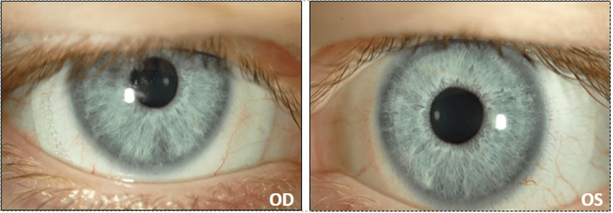
At the patient’s routine follow-up appointment, their chief complaint was that of persistent “blur”, “doubling”, and “ghosting” of vision with their PROSE devices, worst at nighttime and with left worse than right eye.

Entering best corrected visual acuity (BCVA) with PROSE:

OD: 20/40+1 (PH 20/20-2)

OS: 20/50 (PH 20/25+2)

Presenting PROSE device parameters:

OD: 18.5mm OAD/ 8.00 BC/ 3.03 sag/ +2.37 -0.50 x 005/ 0.3 CT/ quadrant-specific design

OS: 18.5mm OAD/ 8.00 BC/ 3.03 sag/ +2.50 -0.75 x 030/ 0.3 CT/ quadrant-specific design

The fit for each PD was found to have adequate corneal and limbal clearance throughout. No edge staining was noted on the conjunctiva after PD removal for either eye; however, there was moderate rebound hyperemia which was concerning for suction with PD wear.

Slit lamp examination (SLE) corneal findings showed mild apical thinning and steepening, with Vogt’s striae and positive presence of a Fleischer ring for each eye. No corneal scarring or neovascularization was noted for either eye. The patient’s intraocular pressure (IOP) with Goldmann applanation tonometry (GAT) was 15 for each eye. All other findings, including pupils, and extraocular motility (EOM) were found to be normal. Posterior segment findings as reported from a recent dilated fundus examination (DFE) from her primary eye care provider, a corneal specialist, showed normal posterior segment findings. The patient’s medical history and systemic medications were noncontributory. They were not using any prescribed or over-the-counter ophthalmic medications.

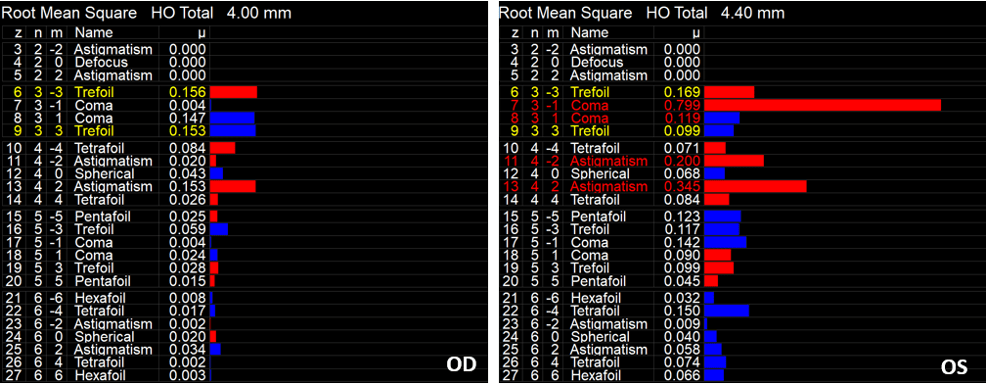
The patient’s visual complaints were consistent with that of residual HOAs. To quantify the degree of HOAs with her presenting PDs, corneal aberrometry scans were obtained for each eye. Aberrometry scans confirmed residual HOAs, left eye worse than right (Figure 1).

HOA Aberrometry Scans

Figure 1: iTrace aberrometry scans demonstrating residual HOAs with patient’s presenting PROSE device for the right (OD) and left (OS) eye. Residual HOAs were more pronounced for the left than right eye, consistent with patient’s chief complaint.

After further discussion of clinical findings, the patient confirmed symptoms of suction with lens wear including aching and soreness as well as rebound redness after lens removal for both eyes. The decision was made to modify the fit for both eyes, with the goals of improving comfort by mitigating suction and optimizing vision by attempting to correct for the residual HOAs.

Refitting/ PROSE Retreatment

With PROSE retreatment, the diameter was increased by 0.5mm for both eyes to provide a wider haptic edge to better distribute the weight of the device for each eye (Figure 2). This was done to reduce the amount of suction with prolonged lens wear. Each lens was also converted from a quadrant-specific haptic design to an eight-meridian haptic design for optimal customization and fitting. With retreatment, the patient’s rebound hyperemia and complaints of discomfort after PD removal were resolved.

HOA Correction in Keratoconus

Figure 2: Finalized PROSE fit for the right (OD) and left eye (OS) in 19.0mm designs.

HOA Correction with PROSE

Once the fit was stable for each eye, we pursued HOA correction. Aberrometry scans over the patient’s optimized PDs were acquired under dilated conditions to better quantify the degree of HOAs in scotopic conditions.

The aberrometry data was incorporated into the PROSE design for each eye. A repeat refraction was performed over the wave-front guided PROSE devices, which was included in the final design. The patient reported significant improvement in daily visual function, with improvement of “ghosting” in the left eye and complete resolution of ghosting for the right eye.

BCVA with final wave-front guided PROSE:

OD: 20/20-1

OS: 20/25-1

Final PD Parameters:

OD: 19.0mm OAD/ 8.00 BC/ 3.03 sag/ +2.00 sph/ wavefront optics/ 0.3 CT/ eight-meridian design

OS: 19.0mm OAD/ 8.00 BC/ 3.03 sag/ +2.50 sph/ wavefront optics/ 0.3 CT/ eight-meridian design

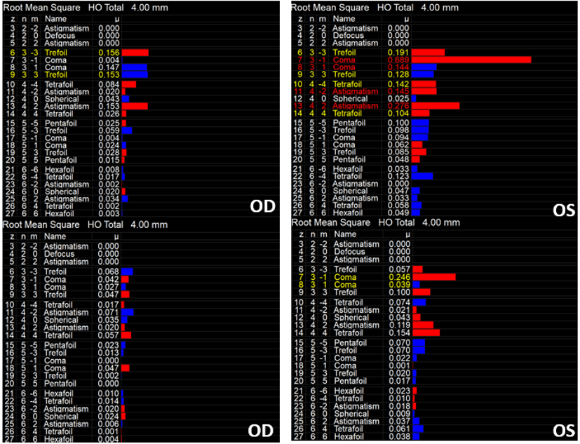
Repeat aberrometry scans were obtained over the final wavefront corrected PDs which confirmed significant reduction in HOAs. The most notable improvement was observed for coma, which can be the most visually significant in KCN patients (Figures 3 and 4).

HOA Correction in Keratoconus

Figure 3: iTrace aberrometry scans with patient’s presenting PROSE devices (top) for the right (OD) and left (OS) eye, compared to patient’s finalized PROSE devices (bottom) for the right (OD) and left (OS) eye.

HOA Correction in Keratoconus

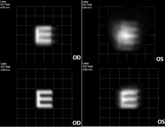
Figure 4: iTrace visual function analyzer (VFA) scan demonstrating the vision quality with the patient’s presenting right (OD) and left (OS) PD (top) versus the patient’s finalized wavefront-guided devices (bottom). There is a notable improvement in quality of vision based on the VFA for each eye.

Discussion

Higher Order Aberrations and Scleral Lenses

Bringing wavefront-guided scleral lenses involves various considerations, including determining who may be an appropriate candidate. The HOA profile of each patient is as unique as their fingerprint – no two eyes are the same. The recipe for HOA correction with scleral lenses has four key ingredients:

  1. Patient must have some vision improvement potential.
    • Improved vision with pinhole.
    • Symptoms of HOAs which can be quantified as HOAs based on aberrometry scans.
  2. Access to an aberrometer, with reliable aberrometry scans.
  3. A rotationally and tangentially stable scleral lens design.
    • Much like the need for a toric soft CL to be stable to function properly, a wavefront-guided scleral lens also needs to be stable while on the eye.
  4. Access to a wavefront-guided lens design with an appropriate manufacturing lab.

Conclusion

Although specialty CLs including scleral lenses can greatly improve visual function and correct for some degrees of HOAs, residual HOAs can haunt patients’ visual function as they experience symptoms like blur, distortion, doubling, shadowing, glare, and ghosting. Correcting for HOAs can further improve visual function in patients suffering from KCN; therein, potentially further reducing the need to pursue invasive corneal surgery such as penetrating keratoplasty.

As technological advances continue to unfold and HOA correction through wavefront-guided technology becomes increasingly available to practitioners, the future is looking increasingly bright for patients.


1. Schornack MM. Scleral lenses: A literature review. Eye Contact Lens. Published online 2015. doi:10.1097/ICL.0000000000000083
2. Sabesan R, Johns L, Tomashevskaya O, Jacobs DS, Rosenthal P, Yoon G. Wavefront-guided scleral lens prosthetic device for keratoconus. Optom Vis Sci. 2013;90(4):314-323. doi:10.1097/OPX.0b013e318288d19c
3. Chalita MR, Xu M, Krueger RR. Correlation of Aberrations with Visual Symptoms Using Wavefront Analysis in Eyes after Laser in situ Keratomileusis. J Refract Surg. 2003;19(6):682-686. doi:10.3928/1081-597X-20031101-13
4. Naderan M, Shoar S, Kamaleddin MA, Rajabi MT, Naderan M, Khodadadi M. Keratoconus Clinical Findings According to Different Classifications. Cornea. Published online 2015. doi:10.1097/ICO.0000000000000537
5. Marsack JD, Ravikumar A, Nguyen C, et al. Wavefront-guided scleral lens correction in keratoconus. Optom Vis Sci. Published online 2014. doi:10.1097/OPX.0000000000000275
6. Koppen C, Kreps EO, Anthonissen L, Van Hoey M, Dhubhghaill SN, Vermeulen L. Scleral Lenses Reduce the Need for Corneal Transplants in Severe Keratoconus. Am J Ophthalmol. Published online 2018. doi:10.1016/j.ajo.2017.10.022
7. Chahal JS, Heur M, Chiu GB. Prosthetic Replacement of the Ocular Surface Ecosystem Scleral Lens Therapy for Exposure Keratopathy. Eye Contact Lens. 2017;43(4):240-244. doi:10.1097/ICL.0000000000000265

Thank you to Bita Asghari for contributing to Global Insight.

[us_separator size=”small” show_line=”1″]
[us_image image=”9240″]
[us_separator size=”huge”]

Dr. Asghari is an optometrist and the Associate Director of Clinical Education for BostonSight. She also serves as adjunct clinical faculty for MCPHS school of Optometry and New England College of Optometry (NECO). She completed her Doctorate in Optometry from Southern California College of Optometry, graduating summa cum laude. Prior to joining BostonSight, Dr. Asghari completed her PROSE clinical fellowship, as well as her post-doctoral residency in Cornea & Contact Lens and Ocular Disease from The Ohio State University Havener Eye Institute.

[us_separator show_line=”1″ size=”small”]
[us_post_navigation in_same_term=”1″]
[us_sharing providers=”facebook,twitter,linkedin” align=”right”]
Tags: , ,

Ortho-k and the Emerging Myopic Child

[us_post_title][us_post_date]

By Martin Conway, Dr Artur Khasanov and Aigul Esmukhambetova

На русском языке

Introduction

Whilst orthokeratology (ortho-k) was originally used as an alternative to spectacles and regular contact lenses and is still a viable option for those candidates, it is used these days predominantly for the management of the progression of myopia in children.

A by-product of compressing the central zone of the cornea in order to correct the existing myopia is that the periphery of the optic zone receives a relative increase in plus power equal to the minus power produced in the centre.

A myope with a correction of -1.00Dioptres (D) will therefore have a +1.00D addition at the periphery of the treatment zone. Likewise, a -3.00D treatment will benefit from +3.00D at the periphery. It is this unintended side effect that gives an ortho-k correction it’s beneficial effect for the young myope.

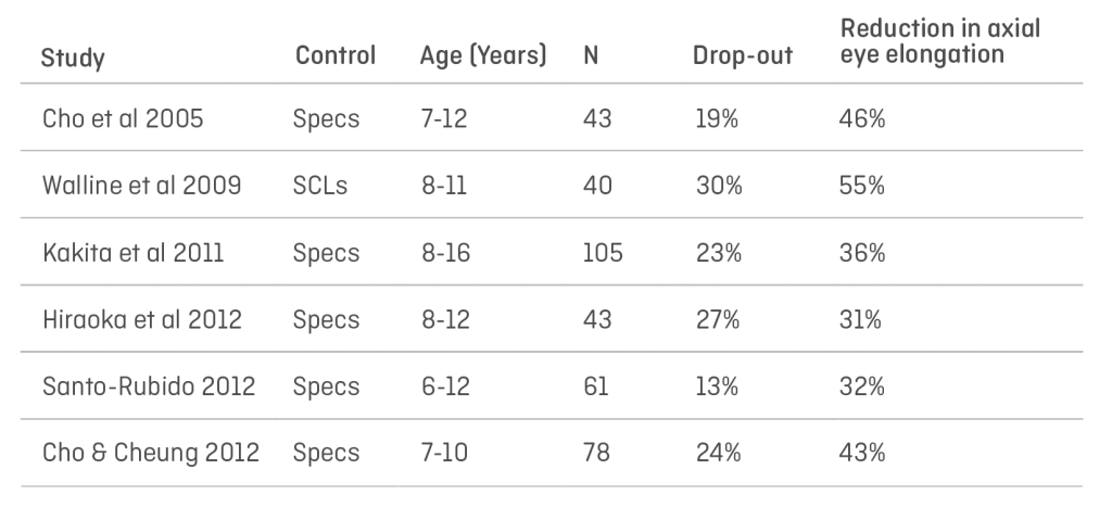
Early studies, looking at the effectivity of standard ortho-k using standard lens designs produced varied results:

effectivity of standard ortho-k using standard lens designs

The studies all used standard lens designs approved within the various jurisdictions where they were conducted. Spherical equivalent powers were between -1.00D and -5.00D, and therefore the plus power achieved at the periphery varied accordingly as a result. It is my contention that this variation in results was produced by the mix of prescriptions within the individual cohorts – those with higher a proportion of high myopes benefitted from higher plus in the peripheries of those treatment zones and therefore more effective reduction in axial growth.

We are no longer constrained by fixed designs, lens design software packages are now available from a variety of sources that will allow the practitioner to have greater control over the prescribed lens for the individual patient’s needs.

The following case study was kindly prepared by Dr Artur Khasanov, eye care practitioner and owner of “Tochnaya Optika Lab”, Orenburg, together with Aigul Esmukhambetova, optometrist, and clearly demonstrates that increased plus power has a greater effect on the slowing of myopic progression in low myopes than conventional ortho-k designs.

Clinical observations of patients, users of Ortho-K lenses

Patient S., 17 years old. He first contacted us in 2014, at the age of 11.

Visual acuity at the time of the visit to a doctor:

OD 0.4 -1.50. = 1.2

OS 0.3 -1.75 = 1.2

The patient was fitted with Ortho-k lenses. We used ortho-k lenses available at that time in our market. Visual acuity while wearing ortho-k lenses:

Visus 0.8 both eyes.

Visual acuity in 2016:

OD 0.1 -2.75 = 1.2

OS 0.1 -2.75 =. 1.2

For two years of using ortho-k lenses (from 2014 to 2016), the progression of myopia was 1.25 D in the right eye and 1.00 D in the left eye.

At that point, we had the opportunity to independently calculate custom-made ortho-k lenses and decided to fit this patient with them.

Visual acuity after fitting: visus OD and OS = 1.2

Since 2016 to date, there has been no myopia progression and the patient continues wearing custom ortho-k lenses.

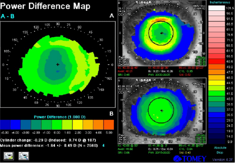
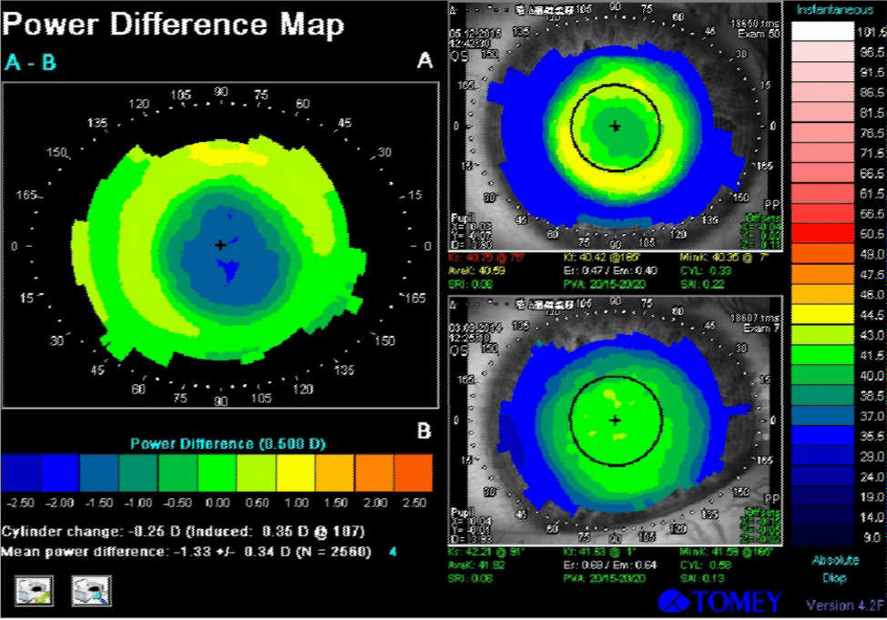
Below are corneal topography images of the right and the left eye during the first lens wear. The tangential power difference map shows that the ring in the projection of the reverse zone is green-yellow. This means that in patients with a low level of myopia it is impossible to achieve a significant myopic defocus on the periphery of the retina. It is in these patients that we observe a more pronounced progression of myopia than in patients with medium and high degrees of myopia.

Ortho-k and the Emerging Myopic Child

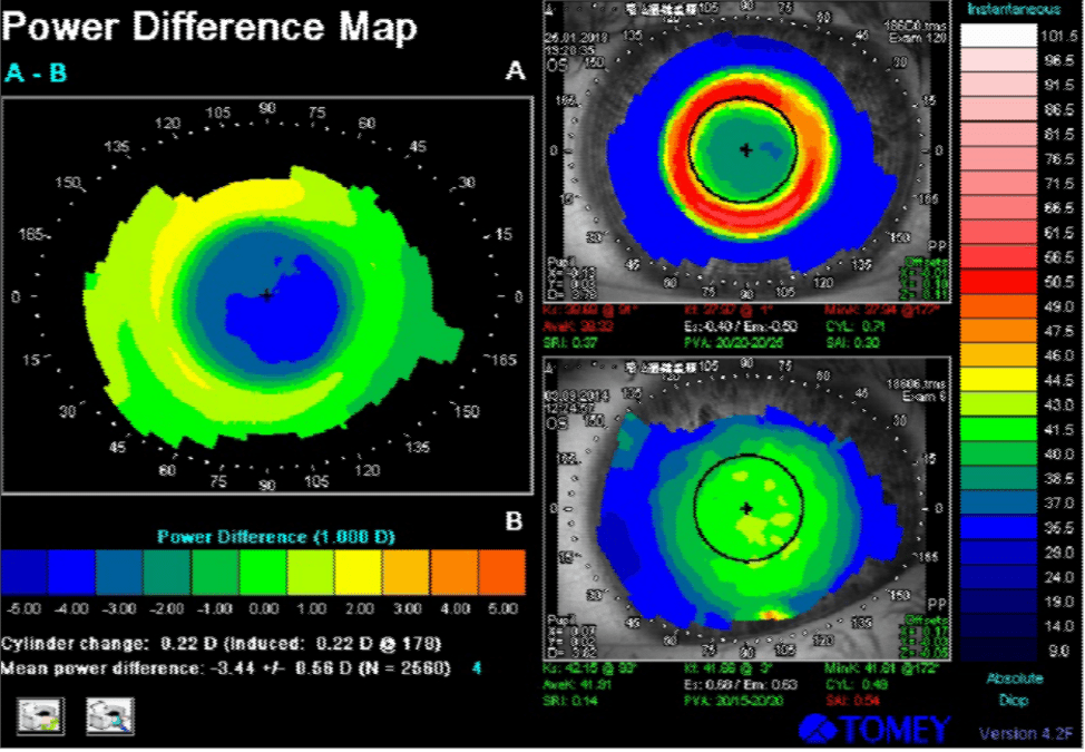
Below are corneal topography images of the same patient, while wearing customised lenses.

Ortho-k and the Emerging Myopic Child

On the corneal topography image, the ring turned red, which means that in the projection of this zone the cornea has become “steeper” and has a high refractive power. We managed to increase the “+” in the projection of the pupil edge and thus achieve a pronounced peripheral myopic defocus.

This was achieved by increasing the tear lens clearance in the lens’ reverse zone. We used increased compression factor and asphericity of the back optic zone. According to our observations, to stabilize myopia, we need to achieve a clearance of at least 80 microns.

Conclusion

Obviously, this is just one example, but results such as these are being found around the globe and are pointing to the fact that “traditional” ortho-k designs may not be exerting sufficient forward movement in the peripheral retinal image shell in lower myopes than is required to achieve the optimal effect on the growth of myopia. This means that for the child with a -1.00D prescription, they will progress, albeit more slowly, until their prescription is such that sufficient peripheral power in the periphery is reached.

More research is needed to establish what the “sweet spot” is, but in the meantime, practitioners may wish to look at what options are available from their laboratories to increase the plus effect in the periphery for these patients.

The Future

Existing lathes are capable of producing these designs as Dr Khasanov’s case study demonstrates. More research is needed to establish whether there is a standard minimum amount of plus power in the periphery required to achieve this stabilising effect or whether an algorithm will be necessary to calculate the optimal lens design for each patient taking into account refraction, both peripheral and central, pupil size, etc. 

We are now told to use the term Myopia “Management” rather than “Control” as we cannot accurately predict the outcome of these treatments. Lens design improvements such as outlined above, coupled with developments in instrumentation to monitor progress and amend designs accordingly should mean that we may soon achieve true control of this growing problem.

Thank you to Dr Artur Khasanov and Aigul Esmukhambetova for contributing to Global Insight.

[us_separator show_line=”1″ size=”small”]
[us_image image=”8848″ has_ratio=”1″]
Martin Conway has over 40 years’ experience in the contact lens field as a qualified Contact Lens Optician. He is registered with the UK General Optical Council on the Speciality Contact Lens Register. Martin is a fellow of the British Contact Lens Association (FBCLA), and The International Association of Contact Lens Educators (FIACLE). He has served in the Professional Services role as an educator and clinical adviser on behalf of both Sauflon and CIBA, and now acts as Professional Services Consultant for Contamac. Martin has lectured extensively in Europe, Asia, Russia, North and South America and the Middle East.
 
[us_separator size=”small”]
[us_image image=”9164″ has_ratio=”1″]

Aigul has been working with the ophthalmologist since 2008. In 2013 she qualified as an optometrist. Aigul specialises in fitting any types of contact lenses.

[us_separator size=”huge”]
[us_separator size=”small”]
[us_image image=”9163″ has_ratio=”1″]

Artur graduated from Orenburg State Medical Academy in 1995. He has been working as an ophthalmologist since 1996. Artur specialises in fitting any types of contact lenses. In 2019 he opened a laboratory for contact lens manufacturing in Orenburg.

[us_separator size=”huge”]
[us_separator show_line=”1″ size=”small”]
[us_post_navigation in_same_term=”1″]
[us_sharing providers=”facebook,twitter,linkedin” align=”right”]

Radial Keratotomy And Reverse Geometry Scleral Lenses

[us_post_title][us_post_date]

by David Foresto and Sandra Au

Case

ME, a 61-year-old lady, was referred to our practice for contact lens correction to improve the vision in the right eye. She had a history of right radial keratotomy (RK) in 1991, right retinal tear in 2015, and bilateral cataract surgery with YAG capsulotomy in 2019.  Levels of refractive error prior to RK and cataract surgery were unknown.

ME reported blurred vision and polyopia in the right eye following cataract surgery even with spectacle correction.

Slit lamp examination showed right radial keratotomy incisions extending into the deep corneal stroma with no signs of corneal oedema or neovascularization. The patient had clear IOLs in both eyes.  She had tilted myopic optic nerves and healthy maculae in both eyes. 

Spectacle refraction was R -1.25/-1.00×180 (6/7.5) and L -0.25 (6/5). 

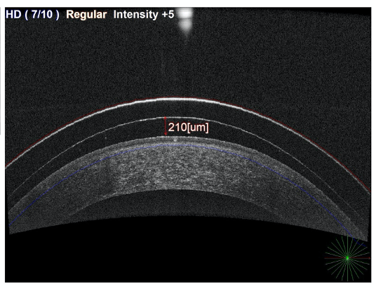
Corneal topography showed a very flat right cornea with mid-peripheral steepening.  K readings were 10.01mm and 10.08mm. Whole eye aberrometry showed medium level higher-order aberrations in the right eye.

Reverse Geometry Scleral Lenses

Refractive correction options were discussed and included updating her spectacles, fitting a soft contact lens or rigid contact lens. We decided to fit her with a rigid contact lens for optimal visual correction in the form of a mini-scleral contact lens to avoid touch to the cornea, improved stability and comfort. She did not require any contact lens for the left eye.

A reverse geometry design scleral trial lens with the following parameters was trialled:

8.28/15.0/+2.50 with Sag 4000 and 2.5D reverse curve.

There was a central clearance of 411um and an over-refraction of -16.50 (6/6-).

The lens was ordered with the following parameters:

9.0/15.0/-8.50 with Sag 3900 and 5D reverse curve.  

The patient achieved 6/6 vision and there was a central clearance of 210um and good limbal clearance. The lens material was Optimum Infinite with Tangible Hydra-PEG surface treatment for supreme oxygen and wettability.

Reverse Geometry Scleral Lenses

ME was very happy with the visual outcome and comfort of the scleral contact lens. She no longer experienced symptoms of blur, haloes or polyopia.

Discussion

Radial keratotomy (RK) was first performed by Russian surgeon Professor Svyatoslav Nikolayevich Fyodorov in 1974 and was popular to correct myopia up until the late 1980s. This surgical technique involved making corneal incisions in a radial pattern, like the spokes of a wheel, to reduce the strength of the peripheral cornea.  The intraocular pressure of the eye would cause steepening of mid-peripheral cornea leading to a relative flattening of the centre of the cornea. The number of incisions could range from 4 to 16, with the centre 4mm of the cornea spared. 

There are various complications associated with RK which can occur early on or late. Complications related to visual outcome include unstable refraction with a tendency to progressive hyperopia, diurnal fluctuation of vision, irregular astigmatism, increased glare and reduced contrast sensitivity. 

Other complications include progressive ectasia, corneal perforations, endothelial damage, increased risk of infection, and incisional neovascularization. Sometimes the complications can lead to the need for a full-thickness corneal transplant.

Often spectacle correction is not adequate due to large anisometropia between eyes, irregular astigmatism, higher-order aberrations, and monocular polyopia.   In the past, hydrogel soft contact lenses were not suitable as they caused hypoxia and incisional neovascularization.  Silicone hydrogel lenses with high oxygen permeability and flatter base curves are sometimes a good option if there is minimal astigmatism and fit the cornea adequately without buckling or fluting.  However diurnal fluctuations in vision may not be corrected by the soft contact lens.

Rigid contact lenses provide best visual outcome and practitioners should generally use a reverse geometry design due to the oblate cornea.  When fitting scleral contact lenses for compromised corneas such as RK, there is further emphasis on ensuring high oxygen transmission to avoid hypoxia and incisional neovascularization. This includes avoiding excessive clearance as a larger tear layer will reduce oxygen transmissibility.  It is important to use a contact lens material which is hyper Dk; the selected material, in this case, was Optimum Infinite, coated with Tangible Hydra-PEG surface treatment. 

Fitting contact lenses for patients with RK can be challenging, however with the development of reverse geometry lens designs and hyper Dk materials, excellent comfort and vision can be achieved without further compromise to the cornea.

Thank you to David Foresto and Sandra Au for contributing to Global Insight.

[us_separator size=”small” show_line=”1″]
[us_image image=”9063″]
[us_separator size=”huge”]

David Foresto is an Australian optometrist and contact lens practitioner specialising in contact lens fitting for keratoconus, corneal grafts and paediatric aphakia. David is a visiting lecturer at the Queensland University of Technology School of Optometry and a former President of Optometry Queensland/Northern Territory.

[us_separator size=”small”]
[us_image image=”9098″]
[us_separator size=”huge”]

Sandra Au graduated from the Queensland University of Technology School of Optometry. She has a special interest in glaucoma and contact lenses, and enjoys mentoring students as a clinical supervisor at the Queensland University of Technology.

[us_separator size=”small” show_line=”1″]
[us_post_navigation in_same_term=”1″]
[us_sharing providers=”facebook,twitter,linkedin” align=”right”]

Keratoconus and Scleral Lenses

[us_post_title][us_post_date]

By Dr Stephanie Woo, O.D.

Patient History

A 34-year-old Hispanic female was referred to us by an ophthalmology clinic for a contact lens evaluation. JS was diagnosed with keratoconus when she was 22 years old and claims that the vision worsened each year. She tried multiple pairs of glasses, and then she was finally fit with a corneal gas permeable contact lens. The corneal gas permeable lens provided good vision, but the discomfort prevented her from wearing it. She then proceeded to have corneal cross linking in each eye in early 2020.

When she saw us for an evaluation, we obtained an in-depth case history. JS told us that she was an LPN (Licensed Practical Nurse) for another doctor in town. Her duties included obtaining case histories from patients, performing tests such as blood pressure, taking temperatures, among others. She also administered injections. JS claimed that she was using the computer for 6+ hours per day. She stated that her very poor vision made it difficult to read the computer or exam notes. As a result, she constantly had to have other people read things for her because she mixed up letters and numbers frequently. Further, she complained of blurred vision, fluctuating vision, double vision, haloes around lights, and eye strain.

Patient Consultation

During her consultation, we performed several tests. The first test was a topography of each eye. The topographical maps provided valuable data so that we could get an idea of how steep and flat her eyes were, along with the overall shape of the cone, the elevation of different parts of her corneas, and her horizontal visible iris diameter (HVID). This information served as a baseline, and more topographies will be taken throughout the course of managing her case over the years.

Topography - OD

Topography – OD

Topography - OS

Topography – OS

The next test was anterior segment photography. These photos provide a documentation of what the ocular structures look like at the time of the fitting. If any scarring, striae or other disorders are seen, photos are taken. This also serves as a good baseline so that if there are any changes in the future, we have something to compare it to.

We then performed a digital retinal wellness scan. This scan checks the macula and optic nerve to ensure there are no retinal abnormalities. This is not totally necessary when fitting a specialty contact lens, but I find it incredibly useful. Many times, if a patient is referred for a contact lens evaluation due to a corneal defect, they end up also having a retinal issue. This is important to discuss with the patient, as their vision potential could be severely limited when they have other ocular abnormalities.

Next, we performed a corneo‐scleral topography. This instrument checks the shape of the sclera. This data is incredibly valuable, as it can tell you the toricity of the sclera and the location of the toricity and elevation. This data helps us select an appropriate scleral lens for a patient. If a patient has a very highly irregular sclera, they may not be a good candidate for a traditional scleral lens. Also, if a patient has a more spherical scleral shape, you may consider a scleral lens that does not have toric haptics.

After all of the data is collected, a slit lamp examination is performed. After, a subjective refraction is attempted to find out the best corrected visual acuity in spectacles.

JS had entering visual acuities of 20/400 in the right eye and 20/400 in the left eye. With a subjective refraction, her vision did not improve in either eye.

Scleral Lens Fit

After reviewing all of her information, we discussed different contact lens options in depth, including corneal gas permeable lenses, hybrid lenses, and scleral lenses. After discussing the pros and cons of each lens, she opted for the scleral lens. Due to finances, she decided to proceed with fitting the right eye only at this time.

Her scleral lens shape showed about 150um of scleral toricity, which is why the Ampleye scleral lens was selected.

The diagnostic lens showed central touch so it was removed and replaced.

The next trial lens showed excessive central clearance, good limbal clearance, and good edge alignment. With an over‐refraction of +9.25, she was able to achieve 20/30 vision!

The final lens was ordered through Art Optical with a decrease in sagittal depth and incorporating the over‐refraction.

  BC Sag Power OAD Toric Haptic
Diagnostic Lens 8.04 4400 -4.00 16.5 150um
Trial Lens 8.04 4800 ‐8.00 16.5 150um
Final Lens 8.04 4600 +2.25 16.5 150um

Lens Material: Optimum Extra

Tint: Clear

Lens Dispense

JS arrived for her lens dispense and we inserted her Ampleye scleral lens in the right eye and she could achieve 20/30 vision immediately. She was thrilled with how well she could see. Central clearance was about 500 um upon insertion. The limbal clearance was adequate and the edges were aligned. 

Keratoconus and Scleral Lenses

Scleral Lens – Central and Inferior – OD

Keratoconus and Scleral Lenses

OCT – Scleral Clearance

We then completed an in-depth training, reviewing lens care and handling techniques. Several devices were used to demonstrate insertion techniques. She was shown 2 different lens care systems to properly clean, disinfect, and store her scleral lens. For scleral lens filling solution, Nutrifill was selected. Nutrifill contains electrolytes – sodium, phosphate, potassium, calcium, and magnesium – to mimic the body’s natural tears and support the ocular surface during prolonged lens wear.

1 day follow up

I called JS the next day to find out how she was doing with her scleral lens. She claimed that everything was in high definition! With her scleral lenses, she stated that she was able to see her computer at work clearly, and she was able to see a menu at a restaurant. She was thrilled with her vision and the comfort of her lens.

3 day follow up

JS called the office complaining that her vision was blurry with the contact lens in. She also stated it was not comfortable. I explained that there was likely an insertion bubble, and she should remove, refill, and reinsert the contact lens. She did as instructed, and was able to see clearly again. Insertion bubbles are one of the most common issues that arise with new scleral lens wearers. It is important to educate them on what to expect when they have a bubble (poor vision, lens discomfort, eye redness, irritation, and foreign body sensation). It is also important that they are instructed to remove the scleral lens, refill and reinsert. Many patients think the bubble will dissipate on its own, and this is not the case. The lens needs to be completely removed from the eye, and the patient should start over.

She will be coming in for her 3 week follow up very soon, and then we will be fitting her left eye into a scleral lens.

Conclusion

Scleral lenses are a viable option for patients with corneal irregularities, such as keratoconus. The gas permeable optics create a smooth surface for refracting light, which results in overall improved visual quality. Keratoconus patients can benefit highly from a gas permeable lens modality and should be considered as a treatment option to improve the vision.

Thank you to Dr Stephanie Woo, O.D. for contributing to Global Insight.

[us_separator show_line=”1″ size=”small”]
[us_image image=”8315″]
[us_separator]

Dr. Stephanie L. Woo graduated Magna Cum Laude from the University of Arizona and graduated with honors from the Southern California College of Optometry. She completed her cornea and contact lens residency at the University of Missouri and specializes in complex corneas and advanced specialty contact lenses. Dr. Woo enjoys lecturing around the world on the topic of specialty lenses and anterior segment eye disease. To date, Dr. Woo has helped over 1,000 patients in the community see better with specialty contact lenses.

[us_separator show_line=”1″]
[us_post_navigation in_same_term=”1″]
[us_sharing providers=”facebook,twitter,linkedin” align=”right”]