By Fareedah Haroun, OD and Chantelle Mundy, OD, FAAO, FSLS
Introduction
Limbal stem cell deficiency (LSCD) results in dysfunction and/or loss of the stem cells in the basal limbal area. These cells are responsible for maintaining and replenishing the corneal epithelium. This change in corneal tissue composition results in conjunctival epithelial ingrowth, persistent epithelial defects, chronic inflammation, and neovascularization.1,5 There are numerous conditions that lead to LSCD, and studies have found that about 15% of cases are a result of contact lens wear.4 Literature suggests that this may be due to mechanical trauma resulting from inadequate lens movement leading to increased friction, compression, and hypoxia. This case report aims to explore the use of scleral lenses to treat LSCD when conventional treatment measures do not improve a patient’s ocular surface.
Case Presentation
A 16-year-old female was referred by the corneal specialist for a scleral lens evaluation due to LSCD of the left eye. The patient participated in a monthly soft contact lens research study for six years and is believed to have developed LSCD secondary to the soft contact lens wear.
Chief complaint: blurred vision, light sensitivity, burning and excessive watering OS
Ocular history: LSCD OS, Myopia OU, Astigmatism OS
Table 1. Entrance Testing and Slit Lamp Examination
Methods and Results
Initial Treatment Plan Prior to Referral:
Discontinue contact lens wear OU
Dry eye management OU
Preservative free artificial tears QID OU
Doxycycline 100mg BID x 7 days
Maxitrol QID OS
Alaway BID OU
Contact Lens Fitting:
OD: discontinue wear of monthly contact lens and refit into daily disposable soft contact lens
OS: fit into BostonSight® SCLERAL 16.5mm with a SmartChannel™
[us_separator size=”small”]
[us_image image=”10945″]
[us_image image=”10946″]
[us_separator size=”small”]
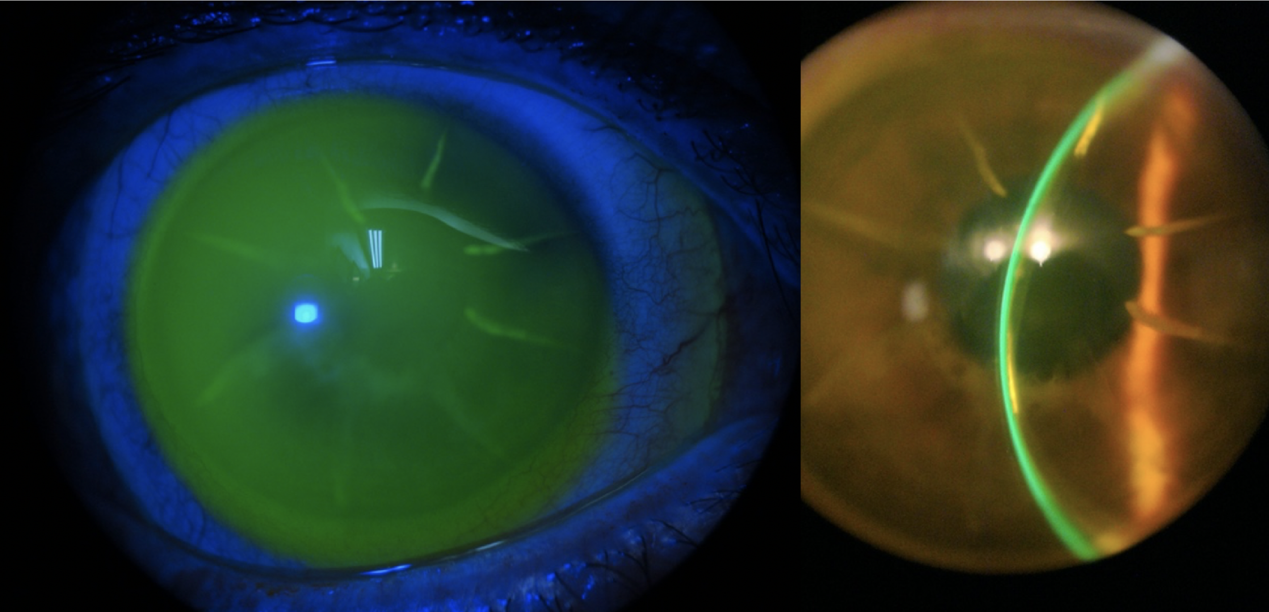
Figure 1. Scheimpflug tomography of the patient’s right and left eye taken prior to contact lens fitting.
[us_separator size=”small”]
Clinical Outcomes:
OD:
BCVA: 20/20
Well centered fit with adequate coverage and movement
No staining upon lens removal
OS:
BCVA: 20/20-
Complete resolution of whorl like peripheral haze after 2 months of wear, residual haze at the superior limbus
No staining upon lens removal
[us_separator size=”small”]
Eye
Brand
Lens
Base Curve
Diameter
Power
Material
Center Thickness
OD
Alcon
Dailies Total 1
8.50
14.1 mm
-4.00 DS
Delefilcon A
n/a
OS
BostonSight®
BostonSight® SCLERAL for Daily Wear
8.00
16.5 mm
-1.48 DS
Contamac Optimum Infinite with Plasma
0.300
Table 2. Final Contact Lens Parameters
Eye
Sphere
Cylinder
Axis
BCVA
OD
-4.25
-0.50
100
20/20
OS
-4.00
-0.75
057
20/20-1
Table 3. Final Manifest Refraction After 5 Months of Scleral Lens Wear
Discussion
Contact lens wear is a lesser known and often asymptomatic cause of LSCD. Most of the published literature suggests that the majority of these cases are due to soft contact lenses. Mechanical trauma is the primary reason for contact lens induced LSCD. This is mainly due to the friction that results as the eyelids push against the ocular surface during a blink. As a result, the eye becomes irritated and dry. If this happens chronically, damage to the limbal area results in stem cell dysfunction. Lens design and materials can also play a role by causing compression and hypoxia at the limbal area.4
Scleral lenses are an excellent management option for LSCD because they provide constant lubrication allowing the cornea to regenerate with minimal disruption. Scleral lenses protect the corneal surface from environmental stressors such as the mechanical trauma that may occur due to the eyelids. Studies have found that the majority of contact lens induced LSCD primarily impacts the superior limbal area. Therefore, superior haptic modifications should be considered to optimize the contact lens fit for these patients.4,5
LSCD can often be misdiagnosed for other conditions. Common misdiagnoses included superior limbic keratoconjunctivitis (SLK), chronic corneal epitheliopathy and corneal intraepithelial neoplasia. Our patient was initially diagnosed with SLK and treated with Maxitrol QID OS. However, SLK is typically bilateral and often has underlying systemic associations.4
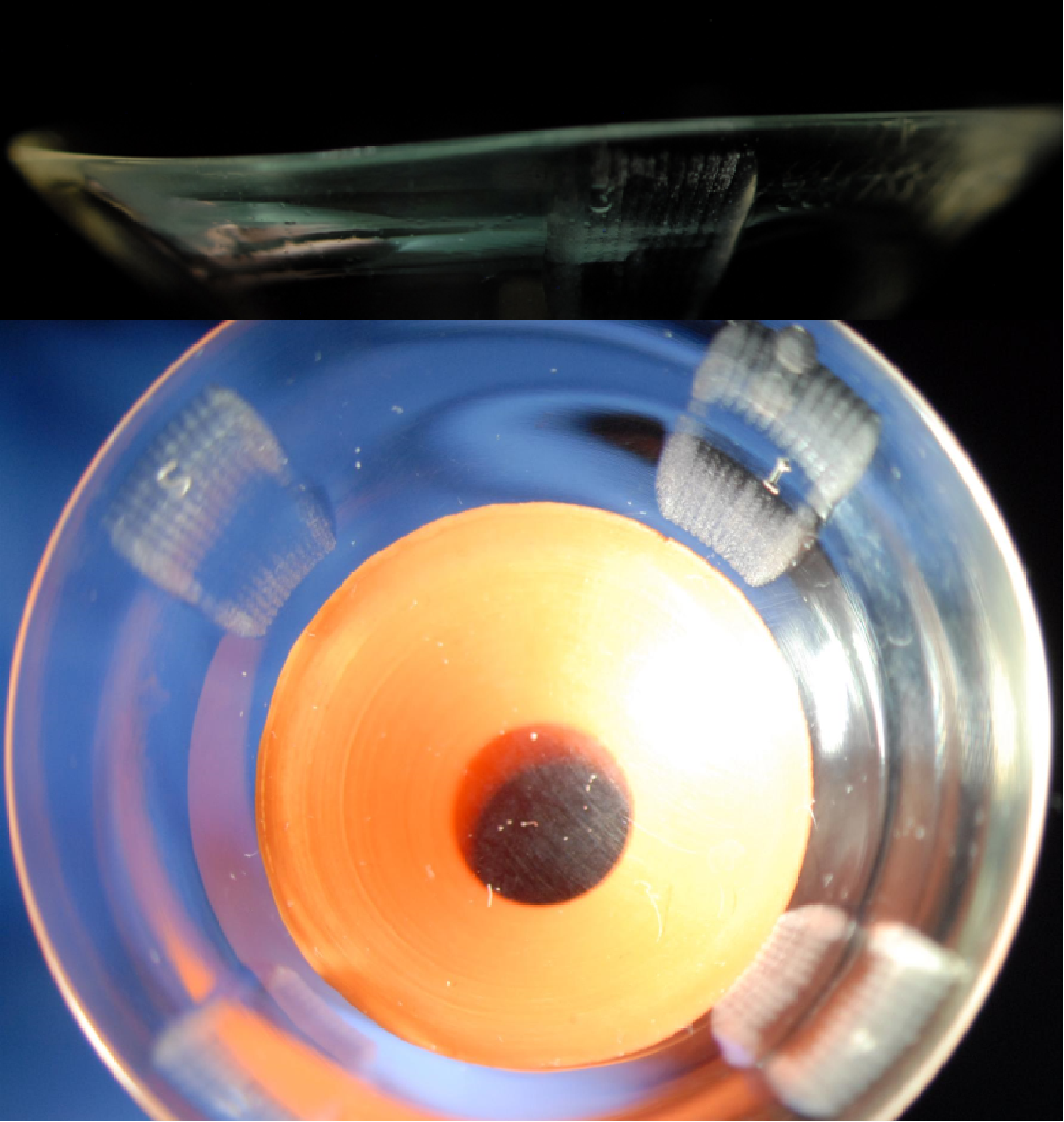
Figure 2. This photo, taken by slit lamp external photography, shows our patient’s left lens at final dispense. The red lines outline the SmartChannel™ that was added to increase oxygenated tear exchange at the superior limbus. The channel is 30 degrees wide with a 0.15um depth.
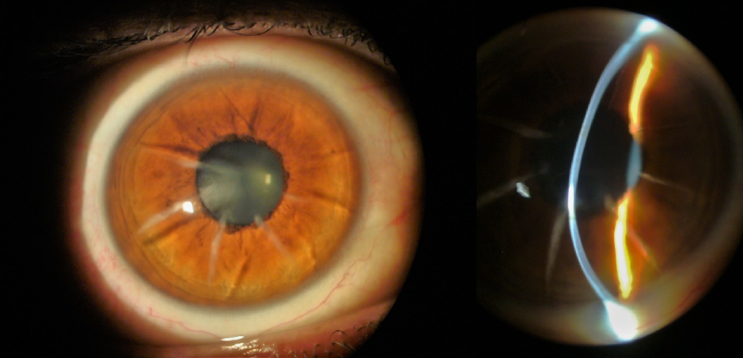
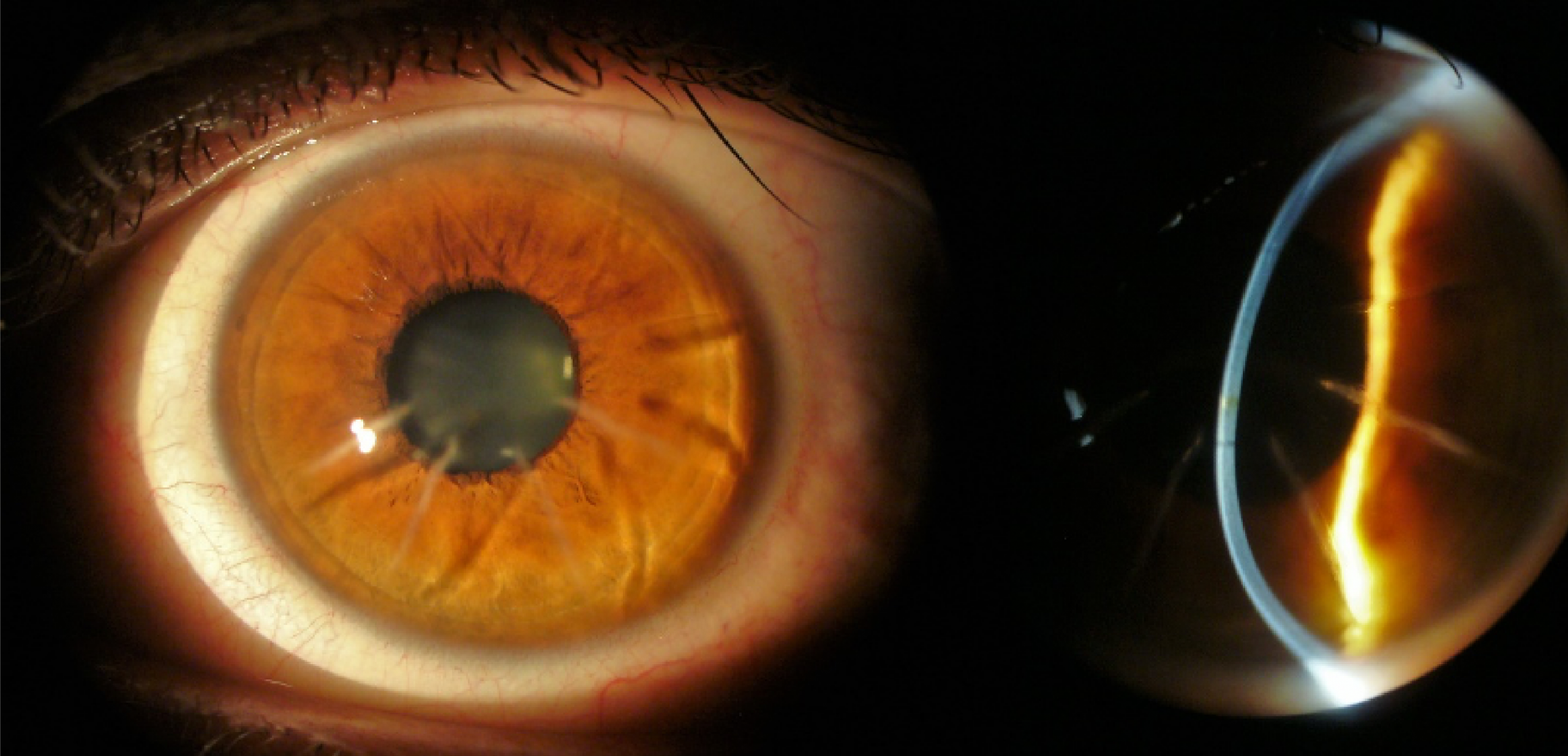
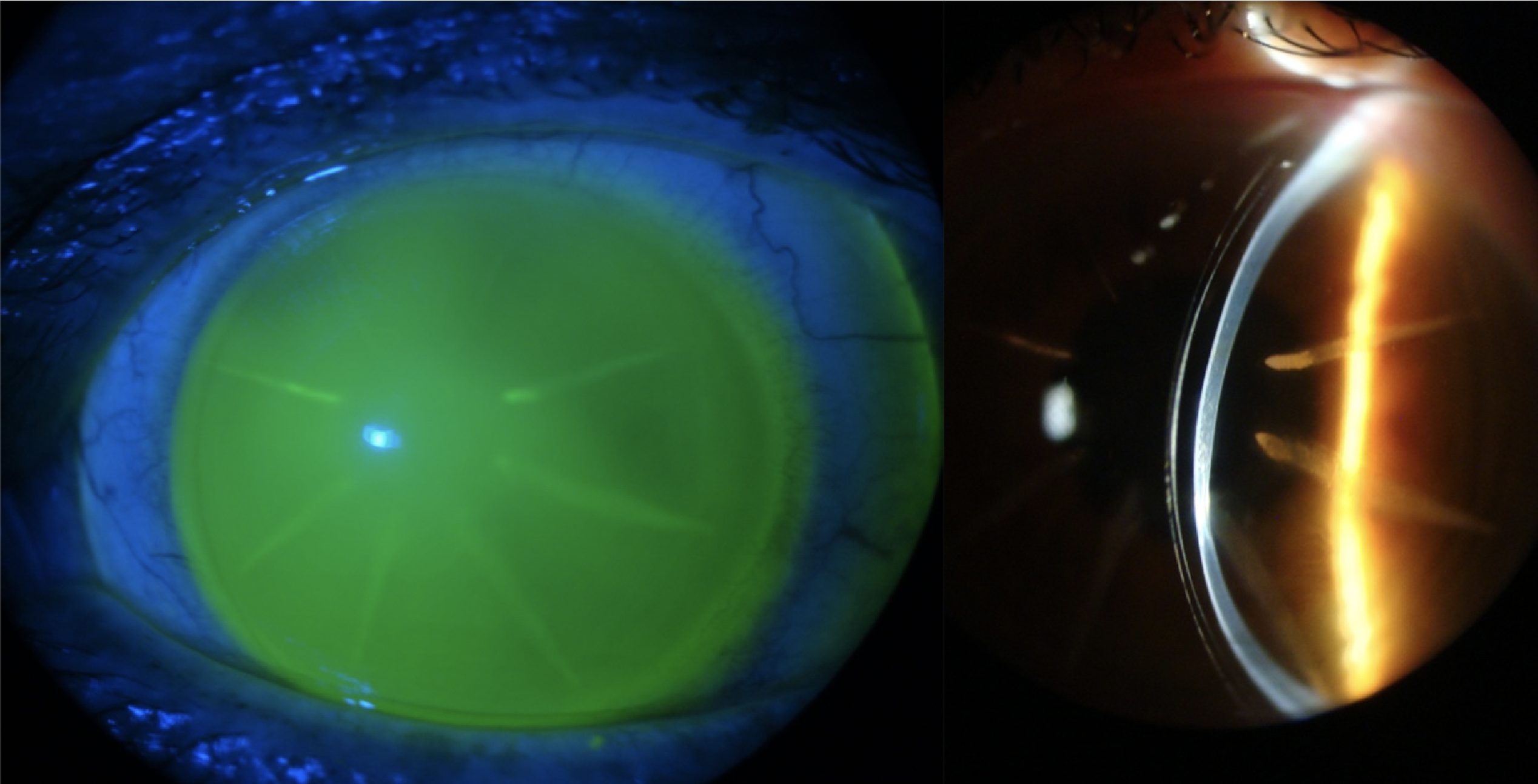
Figure 3. This photo, taken by slit lamp external photography, shows the patient’s ocular surface OS at their most recent follow up appointment. As you can see, the whorl-like fluorescein pattern has completely resolved leaving trace residual haze at the superior limbus.
[us_separator size=”small”]
Conclusion
Contact lens induced LSCD is multifactorial. Contact lenses disrupt the tear film, cause hypoxia and mechanical trauma leading to an inflammatory response.4
LSCD is typically bilateral and asymmetric, but in this case it was unilateral. The patient is at higher risk of developing LSCD in the fellow eye, so you must monitor it closely. To maximize our patient’s ocular health, we switched her into a daily disposable lens and monitored every few months.3
Ensuring that soft contact lenses are properly fit helps to minimize the risk of developing LSCD. As a fitter we should aim for lenses with a low modulus, high DK, and adequate movement (0.1mm-0.4mm). Keep in mind that, although popular, silicone hydrogel lenses have shown increased mechanical trauma due to the decreased flexibility needed to achieve increased oxygen permeability.2
Educate patients on wear schedules, emphasizing no extended or overnight wear. Pay special attention to those with steeper corneas and tighter lids because they are prone to more friction and damage due to inadequate lens movement.4
More conservative treatment options should be explored prior to using scleral lenses to manage LSCD. This typically begins with the cessation of contact lens wear, preservative-free artificial tears, lid hygiene, warm compresses, punctual plugs and other measures as indicated. To address possible inflammation, consider adding topical steroids and/or topical cyclosporine to the treatment plan.4
Surgical management may be considered when all other options are exhausted due to potential risks and complications. Surgical options include, but are not limited to, epithelial debridement, amniotic membrane transplantation and limbal stem cell transplantation.3
[us_separator show_line=”1″ size=”small”]
1. Cheung AY, Sarnicola E, Denny MR, Sepsakos L, Auteri NJ, Holland EJ. Limbal Stem Cell Deficiency: Demographics and Clinical Characteristics of a Large Retrospective Series at a Single Tertiary Referral Center. Cornea. 2021 Dec 1;40(12):1525-1531. 2. Dumbleton, Kathy M.Sc., M.C.Optom, F.A.A.O.. Noninflammatory Silicone Hydrogel Contact Lens Complications. Eye & Contact Lens: Science & Clinical Practice: January 2003 – Volume 29 – Issue 1 – p S186-S189 3. Lee SC, Hyon JY, Jeon HS. Contact Lens Induced Limbal Stem Cell Deficiency: Clinical Features in Korean Patients. Korean J Ophthalmol. 2019 Dec;33(6):500-505 4. Rossen J, Amram A, Milani B, Park D, Harthan J, Joslin C, McMahon T, Djalilian A. Contact Lens-induced Limbal Stem Cell Deficiency. Ocul Surf. 2016 Oct;14(4):419-434.
Dr. Haroun, FAAO, completed her undergraduate degree at the University of Pittsburgh before pursuing her optometric training at The Ohio State University College of Optometry. She stayed in Columbus to complete a residency in Cornea, Contact Lens and Ocular Disease at The Ohio State University Department of Ophthalmology – Havener Eye Insitute. She is now a Clinical Assistant Professor at the University of Pittsburgh Medical Center- Mercy Vision Insitute providing care for patients within the Specialty Contact Lens Clinic.
Dr. Mundy is a clinical associate professor at The Ohio State University Department of Ophthalmology and Visual Sciences. She is a graduate of The Ohio State University College of Optometry and completed an ocular disease-based residency at the Cincinnati Eye Institute. She is program director of the Cornea and Contact Lens residency program and clinical instructor for both optometry students and ophthalmology residents. She is a fellow of the Scleral Lens Education Society (SLS), a fellow in the American Academy of Optometry (AAO) a member of the American Optometric Association (AOA) and completed a PROSE clinical fellowship. Dr. Mundy is co-founder of the Advanced Specialty Contact Lens Clinic in the department and her interests include scleral lens fitting for advanced ocular surface and corneal disease. She received the 2022 Top Doc award from the National Keratoconus Foundation.
Type 1 lattice corneal dystrophy (LCD1) is a rare hereditary bilateral disorder of the cornea. This is a result of an autosomal dominant mutation of the TGFBI (transforming growth factor beta induced) gene which leads to accumulation of amyloid deposits in stroma1
Variable clinical appearance depends on the stage of the disease2
At the onset (usually by age 10): small filamentous anterior stromal lesions, mostly central
With progression: lesions affect posterior stroma and opacify, eventually assuming a ground glass appearance throughout the cornea
By the third decade: most patients will experience recurrent corneal erosions (RCEs) and reduced visual acuity (VA)
Diagnosis:
Biomicroscopy examination alone is often sufficient to diagnose LCD1
Corneal biopsy: can be used for definitive diagnosis3
Treatment: RCE management, phototherapeutic keratectomy and penetrating keratoplasty (PKP) in later stages3
Case Description – Patient MF
64-year-old Caucasian female, diagnosis of LCD1 OU confirmed by corneal biopsy with a corneal specialist.
Past surgical history
The patient had penetrating keratoplasty (PKP) OD in 2016, and cataract extraction (CE) OU (OD 2015, OS 2022).
Chief complaint
MF complained of blurred vision OU with PALs despite PKP OD, in addition to difficulty adjusting to new glasses since OS CE a few months prior.
Habitual glasses Rx with VA
OD -4.75-6.25×075 20/30- (PH 20/20-)
OS plano 20/50 (PH NI)
Add +2.50
Topography
OD tilted corneal graft with inferior elevation and steepening (Figure 1)
OS irregular astigmatism (Figure 2)
[us_separator size=”small”]
[us_image image=”10832″ align=”center”]
[us_image image=”10833″]
Figure 1: Topography of right eye at initial consultation visit
[us_separator]
[us_image image=”10834″]
[us_image image=”10835″]
Figure 2: Topography of left eye at initial consultation visit
Excellent fitting relationship (Figures 5 and 6): central clearance of 210 μm after 4 hours of wear, good mid-peripheral and limbal clearance 360, edges aligned 360.
Patient reported clear and comfortable vision all day OD.
Figure 6: AS-OCT of right eye wearing a scleral lens at two-week follow-up: central clearance of 210 μm after 4 hours of wear
[us_separator size=”small”]
Discussions
For patients with advanced LCD1, scleral lenses can offer superior vision and comfort after PKP. Scleral lenses may improve vision in an eye with corneal dystrophy: significant improvement in BCVA (0.492 logMAR) in a study with 17 eyes with corneal dystrophy.4 However, patient MF was already considering having PKP done on OS before scleral lens fitting, due to having worse BCVA OS post-CE compared to pre-CE. When only one eye has undergone PKP, scleral lens fitting success in the post-operative eye can help accelerate the decision to operate on the other eye.
[us_separator show_line=”1″ size=”small”]
1. Afshari NA, Mullally JE, Afshari MA, Steinert RF, Adamis AP, Azar DT, Talamo JH, Dohlman CH, Dryja TP. Survey of patients with granular, lattice, avellino, and Reis-Bücklers corneal dystrophies for mutations in the BIGH3 and gelsolin genes. Arch Ophthalmol. 2001 Jan;119(1):16-22. PMID: 11146721.
2. Weis JS et al. The IC3D Classification of the Corneal Dystrophies. Cornea Volume 27, Suppl. 2, December 2008
3. Klintworth, Gordon K. Corneal dystrophies. Orphanet journal of rare diseases vol. 4 7. 23 Feb. 2009, doi:10.1186/1750-1172-4-7
4. Parra AS, Roth BM, Nguyen TM, Wang L, Pflugfelder SC, Al-Mohtaseb Z. Assessment of the prosthetic replacement of ocular surface ecosystem (prose) scleral lens on visual acuity for corneal irregularity and ocular surface disease. The Ocular Surface. 2018;16(2):254–8.
Sharon Qiu, OD, MS is a residency-trained optometrist with a passion for specialty contact lenses. She completed an OD/MS dual degree at the New England College of Optometry, where she graduated as the valedictorian of the Class of 2022. After graduation, Dr. Qiu completed a residency in Cornea and Contact Lenses at the University of Waterloo. Dr. Qiu is currently pursuing a full-time PhD in Vision Science at the University of Waterloo Centre for Ocular Research & Education (CORE). She is also serving as a part-time attending optometrist and supervisor for 4th year student interns at the University of Waterloo Contact Lens and Myopia Control Clinic. Her ultimate goal is to become a clinician scientist. She aspires to make a positive impact on her patients’ life, both at an individual level as a clinician, and at a global level as a researcher.
At the core of BostonSight’s mission reside two fundamental pillars: innovation and education. As a non-profit organization, founded in the early 1990s (when there was still so much to learn about scleral lenses, especially after the advent of gas permeable polymers to manufacture these), sharing clinical knowledge and educating about the therapeutic benefits of scleral lenses was, and continues to be, a driving force and guiding light for the organization.
Since the mid-1980s, one of the primary vessels to disseminate our clinical and research data has been via peer-reviewed publications. So much so that BostonSight has been recognized in two bibliometric studies as the number one contributor of scleral lens peer-reviewed publications in the United States, and number two worldwide.1,2 Additionally, for the past 20 years, optometrists and ophthalmologists from all over the globe have come to BostonSight for in-person, immersive education in our own clinic. This has ranged from clinical shadowing to completing the intensive PROSE Fellowship, designed to address the most advanced cases of ocular surface disease. It has also included collaborations with the New England College of Optometry (NECO), MCPHS University College of Optometry, Massachusetts Eye and Ear Infirmary, and the Veteran Affairs Hospital System, all focused on developing and educating future generations of clinicians.
Over the years, our educational evolution and goals have been to broaden the reach, increase the bandwidth, strengthen the impact, foster collaboration, and pass it forward. After almost twenty years of focusing primarily on clinical immersive, in-person education, the concept of FitAcademy™ was born in 2018. It started as a ‘pie in the sky’ concept, then a goal, and later became a reality. Could we transport, host, and “feed” Cornea and Contact Lens residents from all over the United States and Canada at little to no cost to them? Feeding not only their bodies but also their intellect with our 30+ years of knowledge on everything scleral lenses. The content would have a strong emphasis not only on the mechanics of scleral lens design, fitting, and troubleshooting, but an equal emphasis on the importance of management and co-management of complex corneal diseases. The fortune of working alongside corneal specialists throughout the existence of BostonSight and treating some of the most complex and challenging corneal disease patients allowed us to learn early on that to be successful with scleral lenses, practitioners need to see the big picture, need to learn about ocular surface disease management and co-management, need to know how to collaborate and how to communicate with referring specialists, and need to learn how to manage complications beyond the fit. This is how the phrase “it’s not just about the piece of plastic” came to be; coined by our corneal specialist and medical director ca. 2008. Additionally, as pioneers in the field and innovators in lens designs, having acquired a wealth of knowledge on scleral lens design and troubleshooting, it was only fitting (no pun intended!) to share more broadly our knowledge with a wider audience and further our mission of education. And so, FitAcademy™ was launched, expanding our reach to the entire Cornea and Contact Lens Residency classes from the United States and Canada, and just last year to practicing clinicians from Europe (Figure 1).
Figure 1. BostonSight’s educational scope, reach and growth over the last two decades.
It takes a village. Thanks to the generosity of BostonSight donors and individual sponsors like the John Henry Family Foundation, Contamac, Eaglet Eye, Tangible Science, OCULUS, and Glaukos Corporation, this year we are celebrating the 5th anniversary of FitAcademy for Cornea and Contact Lens residents. With its inauguration in 2019 (Figure 2), the hallmark of FitAcademy has always been to involve ophthalmology and external optometrists in its faculty and, in addition to an all-intensive didactic program, include a wet lab with real patients that have a variety of conditions across the entire complex corneal disease spectrum. This model allows practitioners to see the didactic lessons come to life, and through lively discussions and first-hand evaluations of complex patients, enhance their experience and knowledge.
Figure 2. Inaugural FitAcademy for Cornea and Contact Lens residents and fellows – 2019.
Through participant surveys, we continue to strengthen and improve the program and have received phenomenal feedback after every FitAcademy – including our all-virtual program at the height of the pandemic in 2020 (Figure 3).
Figure 3. Social media feedback from attendees after our all-virtual program at the height of the pandemic in 2020.
In 2021, we partnered with NECO who graciously agreed to host FitAcademy at their state-of-the-art Clinical Training Center. Here, practitioners enjoy the benefit of synchronous patient evaluations powered by ION cameras in all the slit-lamps and an area of group teaching and discussion. Practitioners also benefit from the tremendous expertise and depth of knowledge of the NECO faculty (Figure 4).
Figure 4. FitAcademy for Cornea and Contact Lens residents and fellows held at NECO’s Clinical Training Center and in partnership with NECO.
In the summer of 2022, we held our first FitAcademy International, in partnership with OptoAcademy®, an educational organization from Spain. Clinicians from Spain visited our BostonSight headquarters for a two-day program that combined didactic sessions, new patient consultations, patient comprehensive annual evaluations, new patient fittings, and ophthalmology consultations featuring a wide range of corneal diseases (i.e., Keratoglobus, Boston Keratoprosthesis Type 1, Steven’s Johnson Syndrome, Ocular Cicatricial Pemphigoid, and Graft versus Host Disease). Each day culminated with clinical grand round style discussions and practitioners benefitted from workshops in scleral lens edge modifications (Figure 5). After the tremendous feedback received from FitAcademy International, we are hosting our second iteration this summer, anticipating practitioners from Spain, Portugal, and Latin America.
Figure 5. Inaugural FitAcademy International for established practitioners held at BostonSight.
It is our goal to continue to carry out our mission by collaborating with global partners to further scleral lens education and the understanding of corneal disease management and co-management for both optometry and ophthalmology. Our 30+ years of experience with PROSE treatment working alongside cornea specialists has given us a unique perspective on the therapeutic benefits of scleral lenses for ocular surface disease. We have an opportunity to educate ophthalmologists in the hope they will adopt scleral lenses themselves, or refer more patients to their optometry colleagues, opening this treatment option to many patients who may otherwise miss the opportunity.
Thomas Paine once said, “The mind once enlightened cannot again become dark”. FitAcademy programs not only mean to enlighten, but they prevent the mind from becoming dark and aim to turn darkness into light by developing ambassadors of sight. Our ultimate goal: that as many patients in the world that can benefit from scleral lenses, actually do so.
Dr. Karen G. Carrasquillo is Senior Vice President of Clinical and Professional Affairs at BostonSight. In addition, she is an adjunct clinical professor at the New England College of Optometry, an adjunct clinical professor at the school of optometry, MCPHS University, Advisory Board member for the Gas Permeable Lens Institute (GPLI), Fellow of the American Academy of Optometry (FAAO), Fellow of the Educational Society of Scleral Lenses (FSLS), Fellow of the British Contact Lens Association (FBCLA), and Global Ambassador of the BCLA. She is the founder and Program Chair of FitAcademy, an educational retreat in cornea and scleral lenses, and is also a planning committee member for the International Congress of Scleral Contacts (ICSC) Meeting and Global Ophthalmic Women (GLOW) meeting.
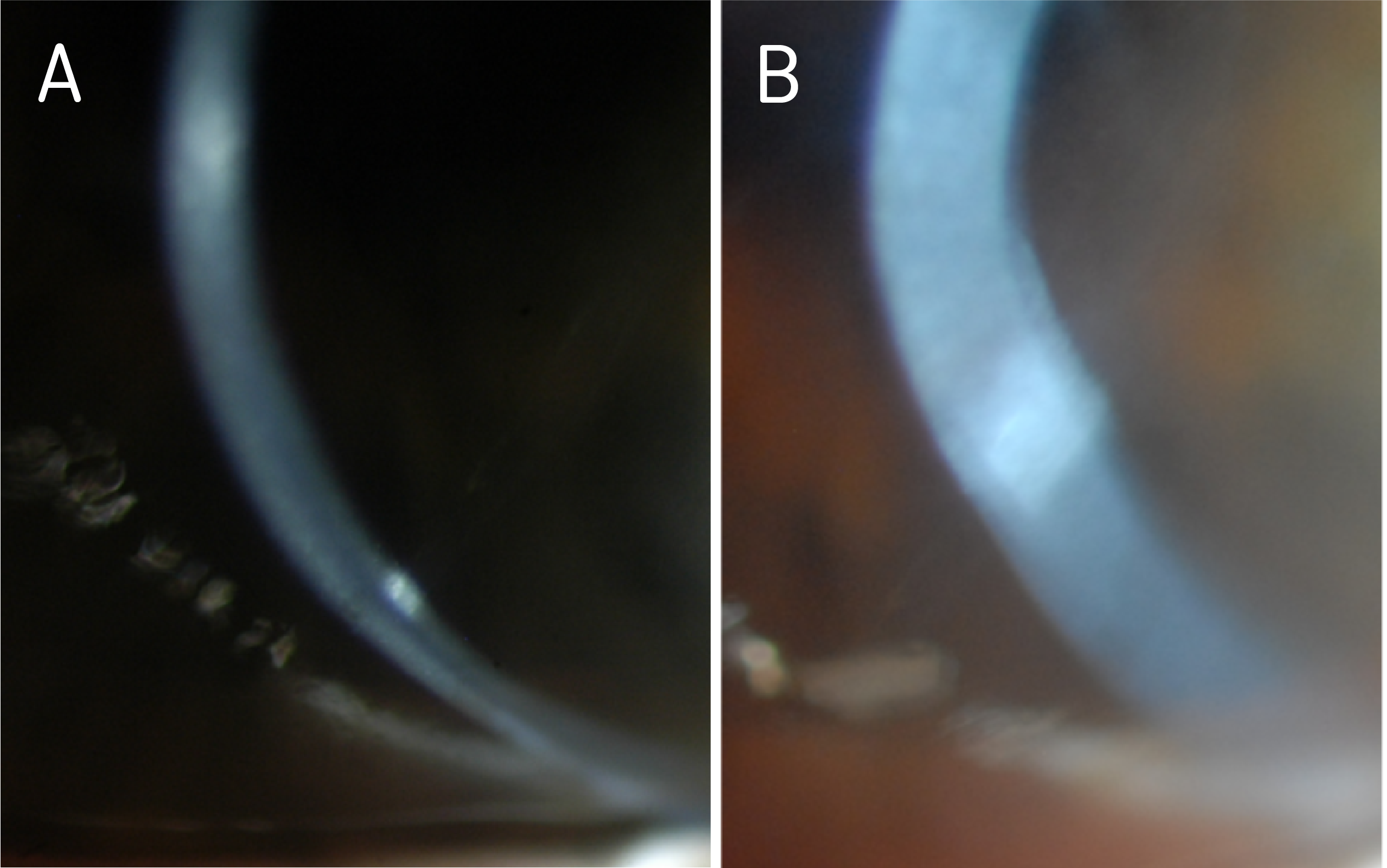
Case of a 60-year-old woman with a history of keratoconus and dry eye syndrome in both eyes, history of corneal scraping in the left eye 25 years prior, history of post hydrops left eye, and questionable potential history of amblyopia left eye. She had a history of successful PROSE treatment since 2010 (11 years of PROSE device wear) but underwent penetrating keratoplasty (PKP) in the left eye in December 2019; secondary to diffuse opacification (Figure 1) and limited best corrected visual acuity.
Prior to PKP in the left eye, she had been wearing 18.5mm, quadrant-specific PROSE devices in both eyes, with added front-surface astigmatism in the right eye and with resultant best corrected visual acuity (BCVA) of 20/30, PH 20/25-2 and 20/100, PH 20/60–, right eye and left eye respectively. With prescribed polycarbonate spectacles to correct residual astigmatism in the left eye and to serve as protection (especially for the better-seeing eye), BCVA in the left eye could be improved to 20/70. The patient wore the spectacles as needed, depending on daily activities and especially during night driving.
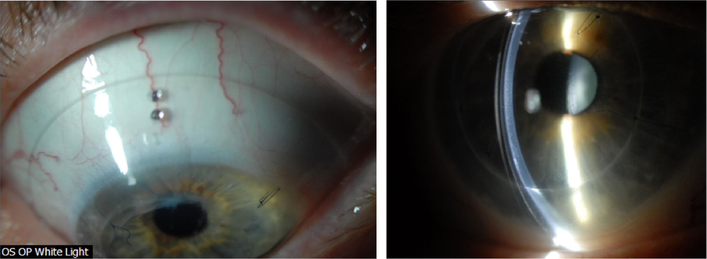
Figure 1. Diffuse haze and opacification in the left eye, as seen on optic section (A) and wide beam (B), prior to PKP.
After corneal transplantation and having been cleared to resume scleral device wear in the left eye, she was referred to our clinic for refitting.
Evaluation After Penetrating Keratoplasty
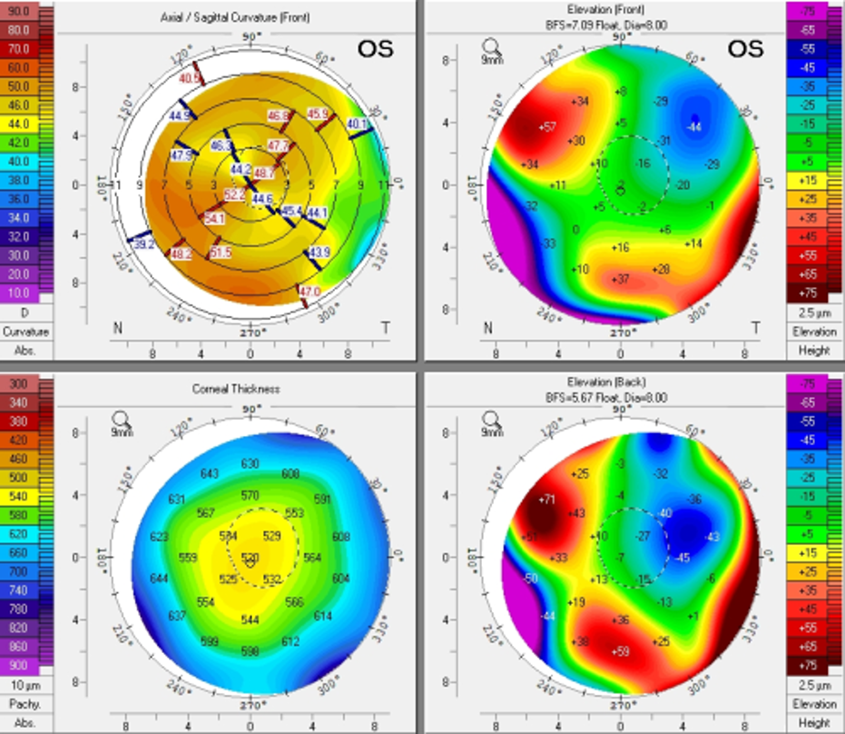
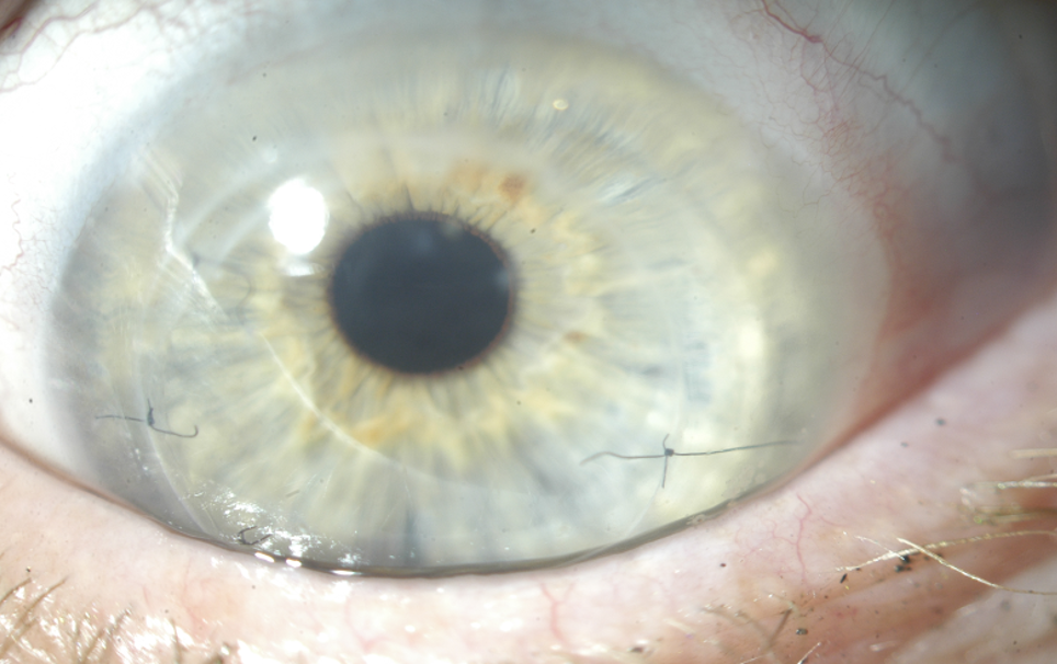
Initial evaluation after PKP OS (Topography shown in Figure 2A), showed a clear and compact graft, with interrupted sutures (Figure 2B). There was no evidence of subepithelial infiltrates, microcystic edema, no keratic precipitates, or stromal diffusion after corneal staining. The patient was on Prednisolone acetate 1% gtts QD and as sutures were removed, she was prescribed Moxifloxacin 0.5% TID in the left eye. Evaluation was also remarkable for cataracts in both eyes, not visually significant at the moment. Best corrected vision during this visit in the right eye was 20/40, PH 20/30-2 and uncorrected visual acuity in the left eye was CF at 3’, PH 20/200.
Figure 2A. Pentacam images s/p PKP in the left eye.
Figure 2B. Presentation of left cornea, status post corneal graft during baseline evaluation for refitting.
Refitting
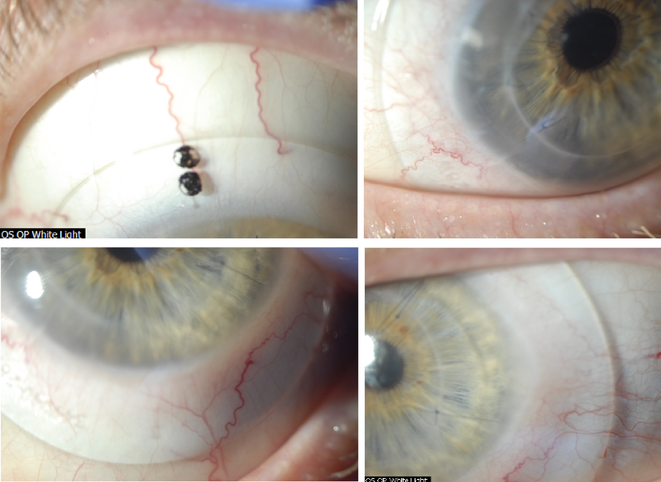
We started a new fit, deciding to keep the patient in the habitual diameter of 18.5mm. Because the cornea was now grafted, we opted to design the lens using Optimum Infinite material (180 Dk) so that together with a center thickness of 0.25mm, oxygen permeability was maximized. Best documented corrected visual acuity in the left eye was 20/30-2 with the new 18.5mm device. While the patient was elated about the newfound visual potential, she reported sensation of pressure during device wear in the left eye – despite no objective evidence of significant compression at the haptics (Figure 3).
Figure 3. Initial fitting endpoint with an 18.5mm PROSE device after PKP OS.
An attempt was made to flatten the haptics, modify the area along the mid-haptic portion to minimize mid-haptic compression and obtain better alignment. Despite these efforts, the patient continued to report pressure sensation with device wear and difficulty with device removal at the end of the day. These symptoms are traditionally associated with lens compression and suction with increased wear time. After reviewing the findings and the thought process with the patient, the decision was made to increase the lens diameter to 19.5mm to provide a larger surface area on which the device could rest. Figure 4 shows the new fitting endpoint with the 19.5mm, 8-meridian quadrant-specific device. One can observe good haptic alignment and the eye is white and quiet, with a nice and even distribution of lens clearance over the grafted cornea.
Figure 4. Fitting endpoint with an 8-meridian, quadrant-specific 19.5mm PROSE device after PKP OS.
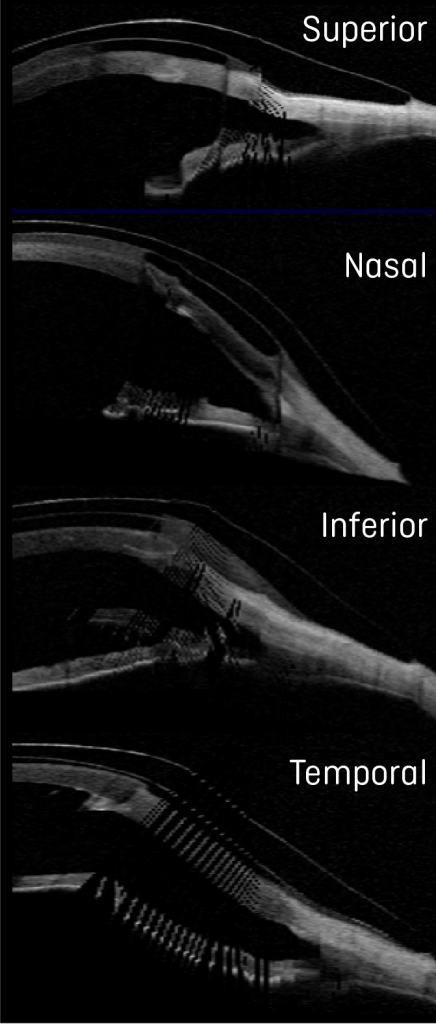
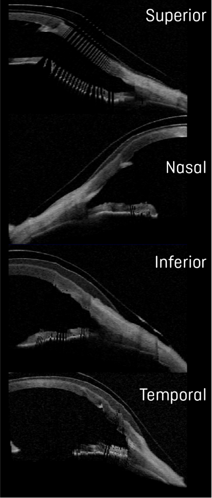
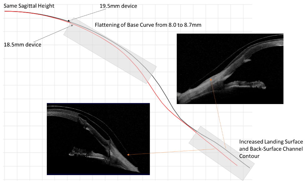
Comparison OCTs show the increased landing surface area when going from an 18.5 (Figure 5) to a 19.5mm device (Figure 6). This attempt to maximize weight distribution over a larger surface area has proven efficacious in our clinical practice when dealing with compression symptoms and device intolerance. While maintaining the same sagittal height, we additionally flattened the base curve to accommodate the more oblate nature of the cornea after transplant as shown in Figure 7.
Figure 5. Ocular Coherence Tomography (OCT) of an 18.5mm device in the different orthogonal meridians of the left eye.
Figure 6. Ocular Coherence Tomography (OCT) of a 19.5mm device in the different orthogonal meridians of the left eye.
Figure 7. Graphical images of 18.5 and 19.5mm, juxtaposed to show the changes to the lens design based on parameter changes using spline curve technology and as observed by OCT. The nasal meridians corresponding to the flattest meridian in the eye are shown for comparison.
Interestingly, while we were optimizing the fit and after reporting improvement in comfort with increased lens size, the vision in the left eye dropped to the 20/50-20/60 range over the course of a couple of months. During all evaluations, we carefully monitored for signs of microcystic epithelial corneal edema, signs of rejection, etc. and at no point were evidence of these found. Given the history of previous cataracts and the fact the patient was on chronic low dose steroids, we performed an undilated assessment of the human lens in the left eye. Photo documentation revealed the development of a posterior subcapsular cataract (PSC) (Figure 8), which was later confirmed to be visually significant during dilated assessment by her corneal specialist. The patient decided to move forward with cataract extraction with hopes to improve vision.
Figure 8. Evidence of PSC formation, during undilated assessment.
Before cataract surgery, we decided to challenge the current 19.5mm design hoping to obtain a comfortable and physiologically compatible lens design. After the patient wore the lens for one week, she reported that the previous symptoms of pressure and difficulty with lens removal had improved, but they were still present. To further minimize compression and suction symptoms, we added 4 back-surface haptic channels to soften further the landing of the haptic surface from bearing harshly on the conjunctiva and prevent a seal off effect (Figure 9).
Figure 9. A 19.5mm, 8-meridian quadrant-specific PROSE device with the addition of 4 back-surface haptic channels to minimize suction and improve comfort.
Discussion and Final Outcomes
Long-term outcomes and benefits of scleral lenses after penetrating keratoplasty have been reported in the literature – while most patients benefit from improvement in visual acuity, complications with scleral lenses, i.e. microcystic corneal edema can ensue in this patient population.1 Special consideration should be taken when fitting or refitting grafted corneas with scleral lenses to minimize the risk of corneal edema, erosions and ultimate rejection. Strategies to promote fluid-ventilation like avoiding tight-fitting edges, lens seal-off and adding back-surface haptic channels can be especially important.1-5 Clinical practice has also shown that the addition of back-surface channels can have the potential to minimize lens suction over time and lead to improvement in physiological endpoints.5-7
In this case, with an increase in scleral lens landing surface area, customized haptic in an 8-meridian quadrant-specific manner and the addition of 4 back-surface haptic channels, not only are we promoting fluid-ventilation to optimize physiological endpoints, but we have a reported significant improvement in comfort. The choice to manufacture lenses in Optimum Infinite material potentiates the oxygen permeability to the grafted cornea. With the final lens after cataract surgery, the patient was able to achieve 20/25+2 best corrected visual acuity with an average comfortable daily wear of up to 12 hours.
By undertaking strategies to progressively soften the landing over the conjunctival surface to reduce or eliminate lens compression over time and minimize suction we were able to achieve a successful refitting outcome.
1. Severinksi, B. et. al. (2014) Scleral contact lenses for visual rehabilitation after penetrating keratoplasty: long-term outcomes. Cont Lens Ant Eye, 37, 196-202. 2. Rosenthal, P. Croteau, A (2005) Fluid-ventilated, gas-permeable scleral contact lens is an effective option for managing severe ocular surface disease and many corneal disorders that would otherwise require penetrating keratoplasty. Eye Cont Lens, 31(3), 130-134. 3. Severinksy, B et. al. (2010) Current applications and efficacy of scleral contact lenses – a retrospective study. J Optom, 3(3), 158-163. 4. Stason WB, Razavi M, Jacobs DS, et al. (2010) Clinical benefits of the Boston ocular surface prosthesis. Am J Ophthalmol, 149, 54–61. 5. Carrasquillo, KG, Byrnes, S (2018) Corneal edema and scleral lenses. Cont Lens Spect, 33, 34-41. 6. Barnett, M (2018) Mind the suction. Scleral Lens Monthly, Nov 2018. 7. Carrasquillo, KG (2020) Mind the suction II. Scleral Lens Monthly, Jul 2020.
Thank you to Dr Karen G.Carrasquillo for contributing to Global Insight.
[us_separator show_line=”1″ size=”small”]
[us_image image=”9526″]
Dr. Karen G. Carrasquillo is Vice President of Clinical and Professional Affairs at BostonSight. In addition, she is an adjunct clinical professor at the New England College of Optometry, an adjunct clinical professor at the school of optometry, MCPHS University, Advisory Board member for the Gas Permeable Lens Institute (GPLI), Fellow of the American Academy of Optometry (FAAO), Fellow of the Educational Society of Scleral Lenses (FSLS), and Fellow of the British Contact Lens Association (FBCLA). She is the founder and Program Chair of FitAcademy, an educational retreat for Cornea and Contact Lens Residents, and is also a planning committee member for the International Congress of Scleral Contacts (ICSC) Meeting and Global Ophthalmic Women (GLOW) meeting.
Patient male, 46 years old, army official, history of post-refractive keratotomy OU performed in 1994, visual acuity was good until 2014 when it began to deteriorate. The patient had to retire early due to his condition and then began to practice law as an army lawyer. He had been wearing glasses which gave poor quality of vision and reduced acuity. He came to us from another state after visiting several doctors and testing different contact lenses in an attempt to improve his vision.
He was fitted with full scleral lenses and the results were an optimal fitting relationship, great comfort and excellent visual acuity while also preserving anterior eye and corneal physiological health.
Introduction
Our clinic has a long history of fitting irregular cornea conditions and since the early introduction of refractive radial keratotomy, many patients were referred to us in order to restore vision in cases where soft lenses or spectacles would not offer satisfactory results. During the early 90’s we relied upon the 12.7 mm wide RGP intra-limbal designs to provide the larger overall diameter to resolve cases like this. It was always a challenging process but our efforts did help many patients.
By 2014 we were able to utilise a full scleral lens design we developed after many years of research into the manufacturing and fitting of scleral lenses. My father, Dr Saul Bastos, MD asked me to study scleral lens technology and suggested that I contact his professor and friend Dr Perry Rosenthal, MD PhD (US) who became my mentor on the subject for years. I also relied on a great friend and specialist in the UK, Dr Ken Pullum FC Optom DipCLP.
During the first experimentations on manufacturing scleral lenses in 2006, we only were able to manufacture up to 16.0 mm. In the following years, new materials and diameters were available which allowed us to manufacture scleral lenses as large as 21.0 mm. There are some cases in which large, full scleral lenses are necessary, especially when there is a disease like dry eye or a large cornea with a large palpebral aperture.
In this case, we faced an irregular corneal condition with a large cornea and palpebral aperture which clearly needed a higher SAG and a wide landing zone. The fact that it was a post-surgical cornea that had been damaged by incisions and now had scars together with reduced mechanical strength due to damage to the collagen fibres also made me aware of the importance of oxygen delivery to the cornea. At the time of the fitting, we were working with the Optimum Extreme (125 DK) from Contamac, it was reassuring to be able to rely on a higher oxygen permeability as this case required full scleral lenses.
Examination
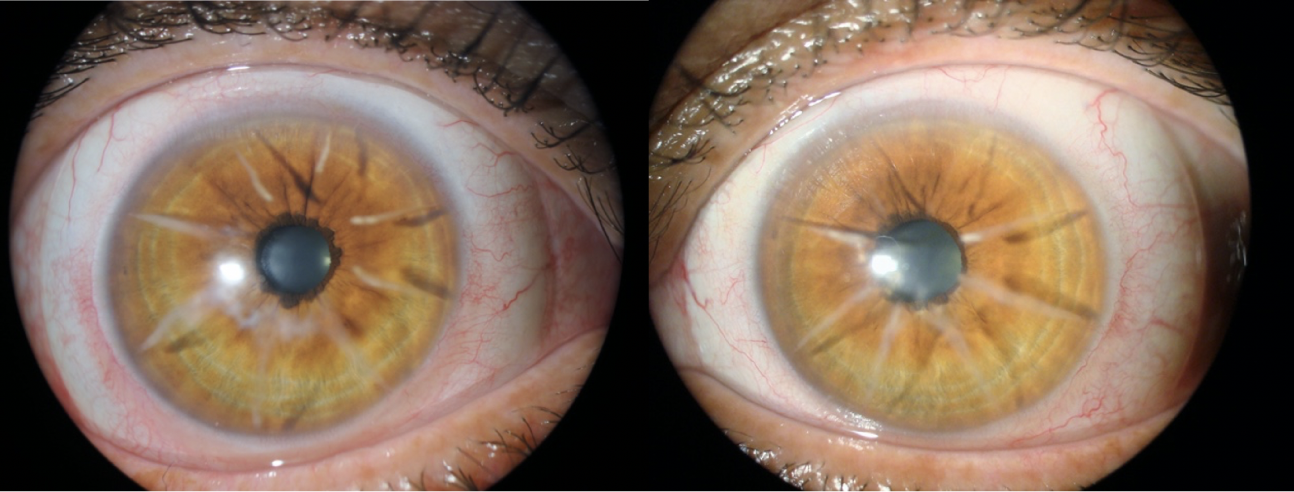
Biomicroscopy showed a series of eight radial incisions with significant depth OU and flatter topography at the central cornea. A decreased corneal thickness was also observed at the central and paracentral cornea. The slit-lamp cross-section view showed an abnormal corneal profile (Figures 1 to 4).
Figures 1 and 2: OD radial incisions and irregular cornea profile under slit-lamp.
Figures 3 and 4: OS radial incisions and irregular cornea profile under slit-lamp.
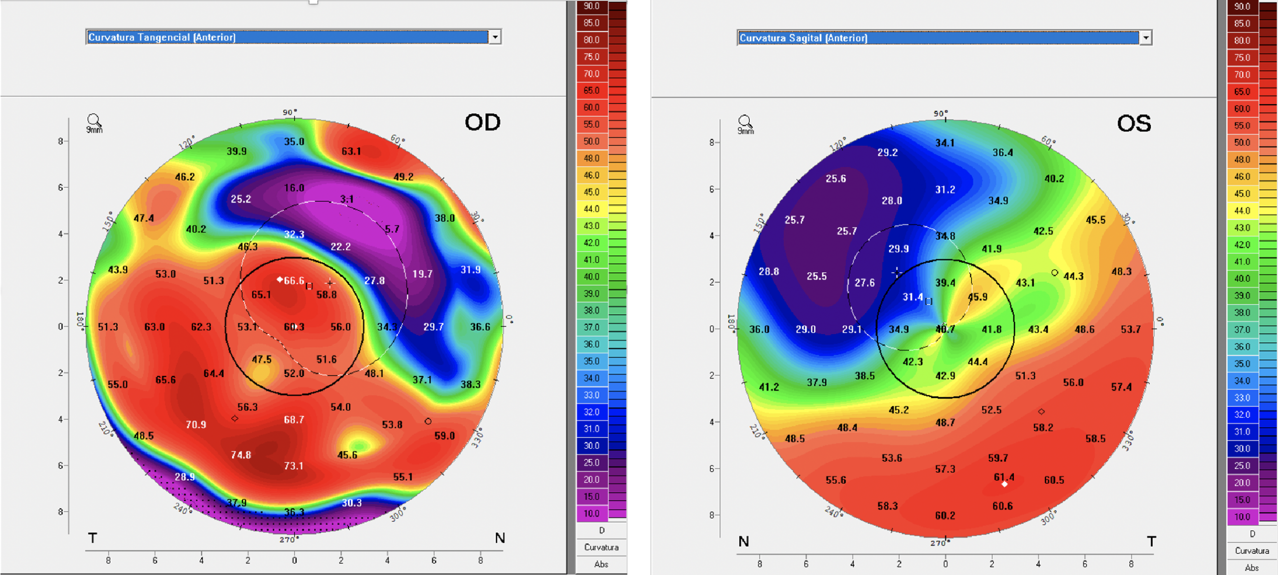
Endothelial cell count was lower than normal in both eyes, worst at OD, so we definitely would need a high Dk, a full scleral lens design and the system I developed called Wave Spline Technology (WST). WST is a ventilation channel system that allows tears to enter the fluid lens reservoir to help to deliver more oxygen to the cornea. We also performed a Pentacam Scan (Figures 5 and 6) and collected data from the precise maps this technology offers together with an OCT to verify each and every quadrant to optimise the landing haptic for a gentle and smooth fitting.
Figures 5 and 6: Anterior Sagittal Curvature large maps.
The patients that submitted to corneal refractive procedures were normally higher myopes and the larger amount of correction required necessitates deeper and more numerous incisions. Back in the day when this procedure was performed, the initial protocol was four radial incisions and, if additional correction was needed, an extra four radial incisions were made. I remember being in an ophthalmology congress with my father, Dr Saul Bastos, at which some surgeons were debating this technique and one of them said that he would perform a total of 12 or more incisions. That was really surprising to my father and me, as we were seeing those patients with those outcomes.
The procedure was abandoned with the advent of laser technology, which also has its complications. The reason I highlighted high myopia is that these patients should be carefully evaluated for glaucoma and retinal diseases as they are more prone to have posterior eye complications. Dr Marcelo Bittencourt MD (IOSB clinical director) examined the patient as a standard procedure with awareness that he was a high myopic patient before refractive surgery.
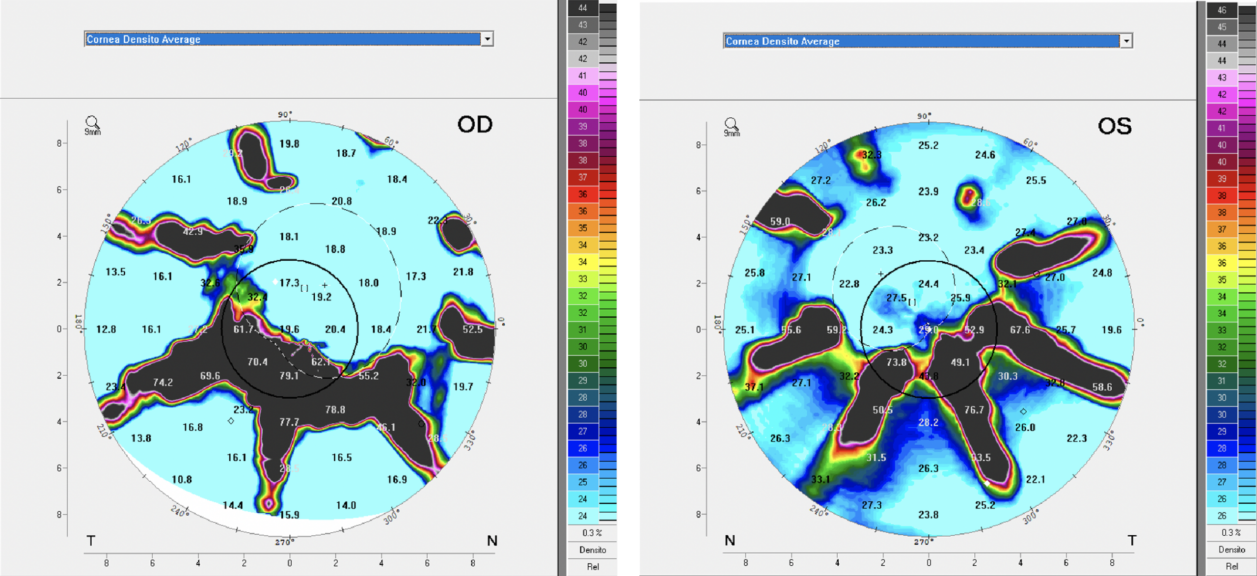
With the Density Average map from Pentacam (Figures 7 and 8), we can also observe the areas of the incisions (in black) with severe opacities. These areas also show the extensions of the incisions and its impact on the corneal epithelium and the corneal topography. The densitometer screening also helps to identify if corneal opacities or haze would change in subsequent examinations.
Figures 7 and 8: Density Average maps identifying opaque areas at the incisions.
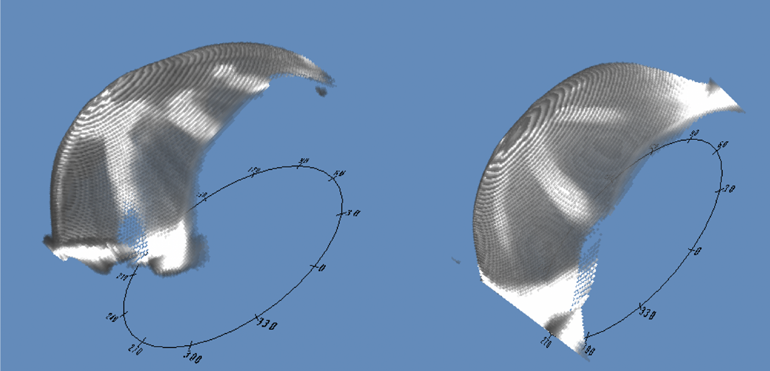
The Scheimpflug and the 3D model images are also great tools to examine abnormal cornea conditions with high irregular astigmatism. The Scheimpflug is similar to the cross-section-view on the slit lamp but it can show the cross-section in different angles. The 3D cornea model is capable of giving a better comprehension of the overall aspect of the anterior cornea and also is capable of showing areas of opacities, it can be rotated in real-time so you can view it from different perspectives, (Figures 9 and 10).
Figures 9 and 10: The 3D Corneal Model images from Pentacam, OD and OS.
We do not hesitate to use all the technology we have to get a comprehensive understanding of the case and data we can use to achieve the best possible speciality lens fit.
Contact Lens Examination and Fit Evaluation
This patient was fitted for the first time in 2014 with 19.6 mm scleral lenses. He wore the lenses for three years with no complications and came for a refit in 2017. Both fits were with the Optimum Extreme material (125 Dk).
In 2019, the patient came for a follow-up and I discussed the new Optimum Infinite material from Contamac that has a Dk of 180. The patient was happy to know more about it, and subsequently, he was refitted with the Optimum Infinite material. The lens parameters were:
OD
OS
BC
47.50 x 45 (7.10 x 7.50)
44.00 (7.67 mm)
Power
-15.00
-8.50
OAD
19.6 mm
19.6 mm
SAG
7.711
7.270
Material
Optimum Infinite
Optimum Infinite
Figures 11 and 12 front images of the model SB Full Scleral lens, OD and OS.
Observe OD the enlarged opacity below the pupil, also visible in the density average map from Pentacam (Figures 7 and 11). The OS image (figure 12) also shows the visible incisions from the refractive keratotomy and it also can be compared to the Density Average image (figure 8) from Pentacam.
Initial thoughts were to design a reverse scleral lens design with a flatter posterior central base curve and a secondary, steeper base curve to overcome the flatter pattern at the central cornea. However, we were able to fit a double base curve, adjusting the base curve radius and optical zone diameters to achieve a good fit on OD as seen in figures 13 and 14.
At OS we designed a single base curve with a large optical zone diameter and we could overcome all the elevations on the cornea (Figures 15 and 16). Both eyes were fitted with a SAG that proved to be high enough after settling with four hours of lens wear. The frontal and cross-section view observed and the slit lamp shows the fluid lens reservoir at both perspectives.
Figures 13 and 14: OD frontal and cross-section view on the slit lamp with fluorescein.
Figures 15 and 16: OS frontal view with fluorescein and cross-section view with white beam.
The patient was happy with the comfort, and visual acuity was 20/10 J1 OU. The visual outcome was much better than expected despite the fact he is 46 years old, (so he should be entering presbyopia soon) he was able to read J1 with no add correction. He was informed about the possible future need of add correction, we determined his dominant eye for possible near vision correction when necessary, utilizing reading glasses, monovision or a multifocal design in the early stages.
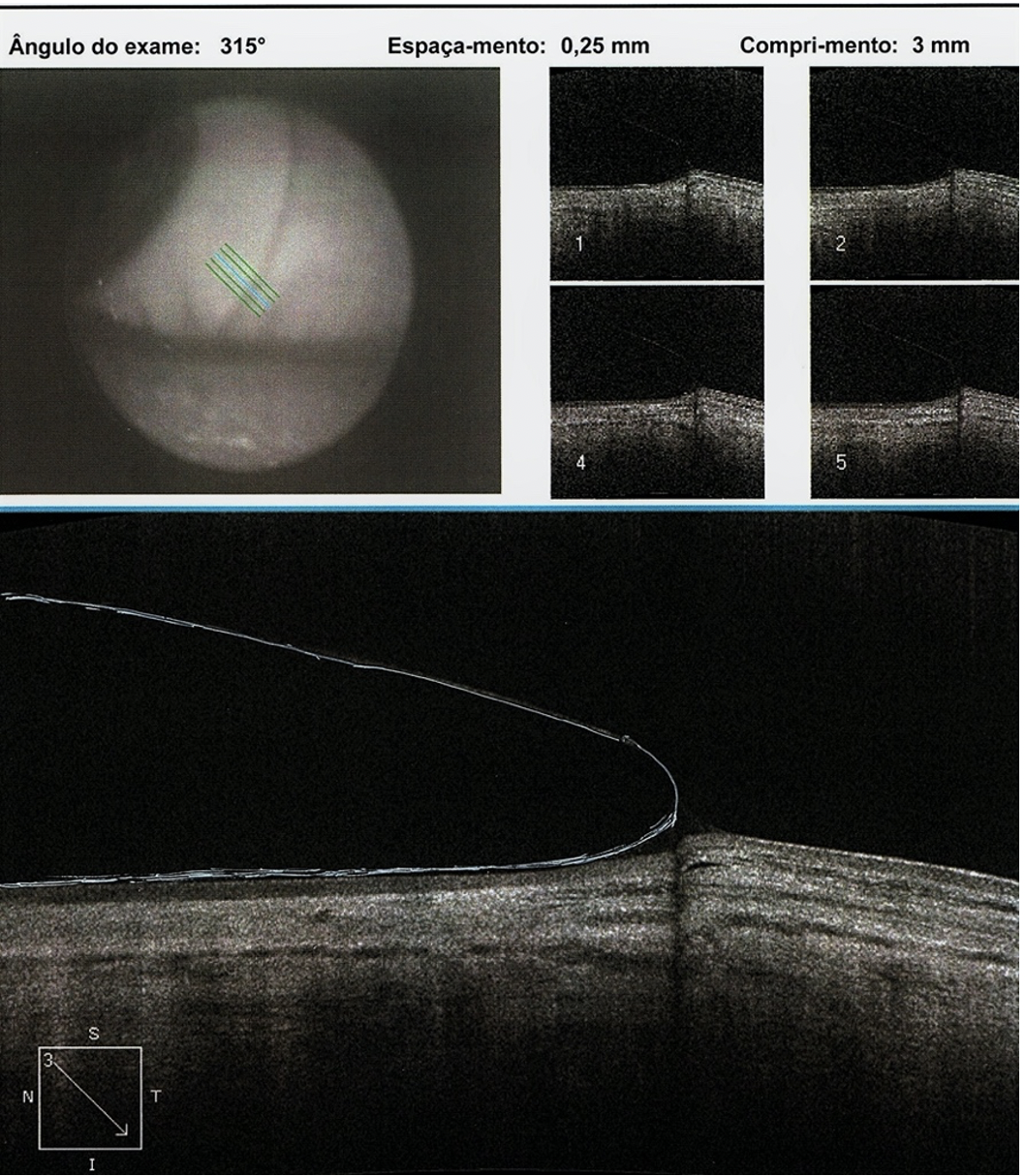
We also performed an OCT scan at eight different angles to have a better understanding of the landing pattern in each meridian. The scans were separated by each angle; one image of the limbal transition and the other of the landing haptic including the edge as you can see in figure 17.
The OCT exam is a great tool to identify areas where the haptic may present significant pressure over the conjunctival sclera, or excessive lift. With all information gathered from these 16 images generated by OCT we can plan and design the scleral lens with an estimation of toricity that we may apply to the haptic and align where required.
Figure 17: A single segment of the haptic showing scleral alignment.
Follow-up Visit After 120 Days
During the weeks following the dispense, the patient reported feeling good, with excellent vision. After about 120 days of dispensing, he was able to visit us again, bearing in mind he lives in another state.
Biomicroscopy during examination showed a crystal-clear cornea, specular biomicroscopy remained stable with a similar endothelial cell count. Visual acuity also remained stable and there was no sign of corneal edema or any complications. Our normal procedure during follow-up visits is to re-evaluate the fitting and consider what might be required to improve the overall fitting. In this case, we decided that with the next lenses we would increase the landing zone and also slightly augment the transitional limbal zone from the base curve to the haptic landing zone. This will allow the lens to have a better pattern over the limbus as it appears to be too close to the limbus (figures 13 and 15).
Customising the Scleral Lens Fitting
When we change any variable at the posterior curve, the optical zones and the overall diameter of the scleral lens SAG are affected. The more variables you change the more impact it has on SAG value, achieving a lower or a higher value depending on the changes made. If you have, like in this case, a SAG that you have as a reference it is a great starting point as you will need to apply the changes to customize the fitting. So, it is necessary to control these variables in a manner that the changes will result in a better fitting relationship while preserving the SAG value to a number close to the reference.
This is absolutely true with trial lenses as well; we always think about customizing the fit whenever it is necessary. If the practitioner does not have software to calculate these changes it is of utmost importance to have technical assistance from a lab consultant that may be able to provide the possible calculations to achieve the results the specialist wants. Another possibility is to use what Ultralentes call Digital Consultancy. In this case, the doctors are encouraged to produce frontal and cross-section view images through the slit-lamp and forward these photos or short videos to the lab. This is often the case as practitioners generally do not use contact lens software to design lenses. Ultralentes also provide the practitioners with a Consultancy Form that the doctor may fill and also make observations and drawings of the specific areas he wants to improve from the trial lens used.
Conclusion
The patient reported feeling really great with his new lenses, he mentioned that he was happy to change to a higher Dk material (Optimum Infinite). He reported greater comfort, visual acuity remained stable and the eye calm. The overall fitting was quite acceptable for a surgical cornea that has multiple incisions that had deteriorated the corneal physiology over the years.
We know that patient’s previous lenses were made in Optimum Extreme and were worn for two years, they were fine and the motive for a new fit was based on taking advantage of higher oxygen transmissibility rather than necessity. Although the lenses were still acceptable to wear, naturally, a new pair may impact visual quality as acuity had no change. What is important is that after four months of wearing the new material, the results were clinically excellent and the patient was happy with comfort and vision.
I also want to address that we had no problem manufacturing these full scleral lens designs in the Optimum Infinite material. It proved to be stable with excellent physical properties with a 180 Dk. This is really important especially when these speciality lens designs are planned.
At the time of writing this case, we already have several scleral lens fittings with Optimum Infinite and it is definitely a great and reliable material from Contamac. The higher Dk material for scleral lens patients means an extended wearing time but is especially important to post-surgery patients.
Here at the Instituto de Olhos Dr Saul Bastos (IOSB), 96% of the fittings are speciality RGP and scleral lens fittings. From these, about 70% are ectasic patients, including (most) keratoconus patients, pellucid Marginal Degeneration and keratoglobus. About 15% are post-surgical patients like post-penetrating keratoplasty (corneal transplants), post-radial keratotomy and post-laser surgery (Lasik and PRK), about 5% are dry eye patients. The remaining 10% of patients are moderate to high myopics, hyperopics, and aphakics. The introduction of the Optimum Infinite material is a great advantage to these post-surgery patients and those with the larger full scleral lens designs.
1. Banta JT. Ocular Trauma – Bascon Plamer Eye Institute. Miami, FL – Saunders Elsevier, 2007 2. Black CJ, Rosenthal P, Kaufman HE et al. Symposium on Contact Lenses. Transactions – New Orleans Academy of Opthalmology. St Louis: Mosby, 1973 3. Bowden TJ. Contact Lens: The Story – Cornwall, UK. London: Bower House, 2009 4. Cochet P, Amiard H. Contact Lens International Clinics. Boston: Little, Brown, 1969 5. Cvintal T, Munarin MAM, Moro F. Complicações do Transplante de Córnea. São Paulo, Santos, 2004 6. Duker JS, Macsal MS, Fontes BM. Anterior Segment – Rapid Diagnosis in Ophthalmology, St. Louis, Mosby Elsevier, 2008 7. Girard LJ, Soper JW, Gunn C. Corneal and Scleral Contact Lenses. Proceedings of the International Congress. St. Louis: Mosby, 1967 8. Girard LJ, Soper JW, Sampson WG. Corneal Contact Lenses. St. Louis: Mosby Co. 1970 9. Godinho C, Dantas B, Sobrinho M.et al. O Padrão CG em Lentes de Contato. Bastos, L. Cultura Médica;. pág. 301 a 313. 2010 10. Jenkin L, Tyler-Jones R. Theory and Practice of Contact Lens Fitting: Hatton Press 1969 11. Pena A. Bastos, S et all, Clínica de Lentes de Contato. Cultura Médica, 1989; pág. 69-79 12. Philips AJ, Speedwell L, Morris J. Contact Lenses – 5th ed. Ed Butterworth Heinmann Elsevier 2007 13. Ferrara, Paulo. Anel de Ferrara – Cultura Médica – 2006 14. Romero-Rangel T, Stavrou P, Cotter J et all. Gas Permeable Ocular Prosthesis Therapy in Ocular Surface Disease. Am J Ophthalmology, 2000, 130: 25-32 15. Rosenthal P, Cotter JM, Treatment of Persistent Epithelial Defect with extended wear of a fluid-ventilated gas permeable scleral contact lens Am J, Opthalm 2000; 130: 25-32 16. Rosenthal P., Cotter JM. The Boston Ocular Surface Prosthesis in the management of Severe Ocular Surface Disease. In: Foulks G. (ed.) Ophthalm Clin A Am 2003; 16: 89-93 17. Schein OD, Rosenthal P, Ducharme C. A Gas Permeable Scleral Lens for visual rehabilitation. Am J Ophthalmol, 1990; 109:318 18. Fadel, Daddi DOptom, FSLS, FBCLA, FAAO – Scleral Lens Issues and Complications: Their Recognition, Etiology and Management. – Dougmar Publishing Group Inc. August 2020
Thank you to Dr Luciano Bastos for contributing to Global Insight.
[us_separator size=”small” show_line=”1″]
[us_image image=”9427″]
[us_separator]
Mr. Bastos is the director and clinical instructor of specialty contact lenses at the Instituto de Olhos Dr. Saul Bastos (IOSB), and is the director and specialty lens consultant of Ultralentes, a small laboratory specializing in GP and scleral lens designs in Porto Alegre, RS, Brazil.
The Scleral Lens Education Society (SLS) is a non-profit educational society whose mission is to teach contact lens practitioners the art and science of prescribing scleral lenses. The SLS provides written, lecture, and video materials on scleral lenses, supporting public education that highlights the importance of scleral lens wear and education. In addition to providing education to practitioners, students, and patients, the SLS fosters a Fellowship program to build and grow the community of practitioners around the world, and is making strides to educate the public and regulatory agencies about the benefits of scleral lenses. The SLS is the go-to resource for all things scleral lenses.
SLS Education
Resources & Materials
One of the primary roles of the SLS is to provide education to practitioners and students. This is accomplished through live lectures and workshops, virtual webinars and workshops, as well as written materials available on our website. All of our virtual education is recorded and available in the living library, among other educational materials. We have resources for students and beginning practitioners, also providing advanced education for experienced practitioners. We also have a popular YouTube channel that has content for both practitioners and patients, including “how to” videos on handling and applying/removing scleral lenses. In addition to educational materials, we have several templates and protocols for scleral lens management, also available on our website in the clinical resources section.
Educational Credit
As a non-profit educational organization based in the United States, the SLS is committed to providing non-biased scleral lens education that is top calibre. We have long been approved to offer COPE credit for practitioners in the United States who are seeking educational credit for licensure. In 2021 we started offering NCLE approval for our events so that our industry partners can also receive credit for education. We have a presence at most US-domestic and an increasing number of international conferences and events, facilitating workshops (Figure 1), providing lectures, and even hosting social gatherings for our Fellows.
Figure 1: SLS board members present the live demonstration-based workshop presented at GSLS 2020. (Pictured from left to right: Drs. Matt Kauffman, Dan Fuller, Pam Satjawatcharaphong)
Patient Education
The SLS website has plenty of education for patients as well, and this is an area we are intent on growing! Currently, our website has a patient site with instructional videos, information on scleral lens indications, as well as a specialist locator that patients can use to find a doctor near them. The SLS is supportive of all public health initiatives and has recently begun to make efforts to teach the public as well as legislative organizations about the benefits of scleral lenses. By explaining the indications for scleral lens use and how to safely handle and care for scleral lenses, we better inform potential patients as well as governing agencies.
SLS Fellowship
The SLS is more than a society to disseminate information, and also works to enhance communication and collaboration among specialists in the field. Our fellowship program allows clinicians and researchers who specialize in scleral lenses to apply for fellowship status. The fellowship process includes submitting lectures, case reports, abstracts, and other items that established expertise with scleral lenses.
Once accepted, these fellows are part of our growing community of educators, clinicians, and researchers that communicate with one another and grow this field so that as many patients can benefit from scleral lenses as possible. Fellows have the benefit of being listed in our “find a specialist” map function for patients, and are also invited to socialize and network at our annual Fellow appreciation event at the Global Specialty Lens Symposium each year. One talented specialist will also receive our prestigious “SLS Fellow of the Year” Award, presented each year at this event (Figure 2).
Figure 2: Presentation of the SLS Fellow of the Year at the annual Fellow’s Reception at GSLS 2020. (Pictured from left to right: Drs. Pam Satjawatcharaphong, Lynette Johns (Fellow of the Year 2020), Karen Lee)
So check out our YouTube channel, visit our booth at the next (live) conference, attend one of our live or online workshops, and visit our website today to learn more about the SLS and how you can get involved! At the very least, become a member today (it’s free!) to get access to all the resources we have to offer.
Thank you to Maria Walker for contributing to Global Insight.
[us_separator size=”small” show_line=”1″]
[us_image image=”9265″ size=”us_600_600_crop”]
Dr. Maria Walker graduated from The New England College of Optometry with an OD/MS degree in 2013 and completed a Residency in Cornea & Contact Lenses at Pacific University in Portland, OR. She then joined the clinical faculty at the University of Houston College of Optometry and is currently part-time faculty while also completing her PhD, studying the effects of scleral lenses on the ocular surface. Dr. Walker lectures domestically and internationally on scleral lenses and is involved in several research projects at the Ocular Surface Institute and Texas Eye Research and Technology Center at the University of Houston. She has won several awards related to contact lenses and research and is a fellow of the American Academy of Optometry, a fellow and the current President of the Scleral Lens Education Society, and an advisory board member of the Gas Permeable Lens Institute.
Scleral lenses have an astounding ability to provide patients with the gift of sight, often allowing them to regain their lives. With keratoconus (KCN) being the most common indication for scleral lenses, patients often experience a significant improvement in visual function with wear.1
It is well-understood that, by providing a smooth refractive surface, scleral lenses have the capacity to correct for visual distortions caused by corneal irregularity. Scleral lenses also correct for some degrees of higher order aberrations (HOAs); however, patients may continue to experience visual distortions from residual HOAs. A patient who continues to be affected by HOAs with scleral lens wear may report symptoms of “ghosting”, “shadowing”, or “doubling” of their vision.2–4 Wavefront-guided correction allows for customized optics at the front surface of contact lenses (CLs), including scleral lenses to correct for a patient’s specific HOA profile.2
Prosthetic replacement of the ocular surface ecosystem (PROSE) is a treatment that uses a highly customizable scleral lens, deemed a prosthetic device (PD), for the management of irregular corneal shape and ocular surface disorders. The PD used in PROSE treatment functions like that of a scleral lens, as it is made of a clear rigid-gas permeable material that is filled with preservative-free saline and applied to the ocular surface and rests on the sclera. With PROSE, customizability in correction for HOAs through wavefront-guided optics is possible. Other scleral lens designs have demonstrated capability to offer wavefront-guided correction as well.5
With the exponential growth of scleral lens wear and technological advances in the industry in recent years, there has been a reduced need for corneal transplants.6 As HOA correction with scleral lenses through wavefront-guided optics becomes increasingly available, practitioners will be able to offer enhanced visual function, which could potentially further reduce the need for corneal transplants in the KCN population.
This report reviews the case of a patient with keratoconus, who had limitations in vision due to residual HOAs. Wavefront-guided optics corrected for their residual HOAs, which significantly improved the patient’s visual function and deterred their interest in pursuing invasive corneal transplant surgery.
Case Report
Patient History
A 54-year-old Caucasian with ocular history of bilateral KCN presented to BostonSight for follow-up. They initially underwent PROSE treatment in 2017 and continued with daily wear of their PROSE devices for visual function. Prior to wearing their PROSE devices, they had tried and failed with all varying types of CL modalities including corneal gas permeable (GP) lenses, hybrid CLs, and other scleral lenses.
The patient’s profession in health care and management had a high vision demand. They were motivated to better their vision to improve their daily visual function. The patient had been advised in the past that should vision improvement be limited with specialty lenses, they may have to consider a corneal transplant. Although this was a notion which the patient was able to forgo for years since their diagnosis, they anticipated that this may be the next appropriate step in their vision journey to restore their visual function so they may meet their daily visual demands.
Case Presentation
At the patient’s routine follow-up appointment, their chief complaint was that of persistent “blur”, “doubling”, and “ghosting” of vision with their PROSE devices, worst at nighttime and with left worse than right eye.
Entering best corrected visual acuity (BCVA) with PROSE:
The fit for each PD was found to have adequate corneal and limbal clearance throughout. No edge staining was noted on the conjunctiva after PD removal for either eye; however, there was moderate rebound hyperemia which was concerning for suction with PD wear.
Slit lamp examination (SLE) corneal findings showed mild apical thinning and steepening, with Vogt’s striae and positive presence of a Fleischer ring for each eye. No corneal scarring or neovascularization was noted for either eye. The patient’s intraocular pressure (IOP) with Goldmann applanation tonometry (GAT) was 15 for each eye. All other findings, including pupils, and extraocular motility (EOM) were found to be normal. Posterior segment findings as reported from a recent dilated fundus examination (DFE) from her primary eye care provider, a corneal specialist, showed normal posterior segment findings. The patient’s medical history and systemic medications were noncontributory. They were not using any prescribed or over-the-counter ophthalmic medications.
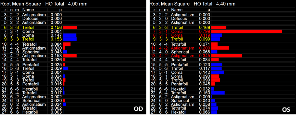
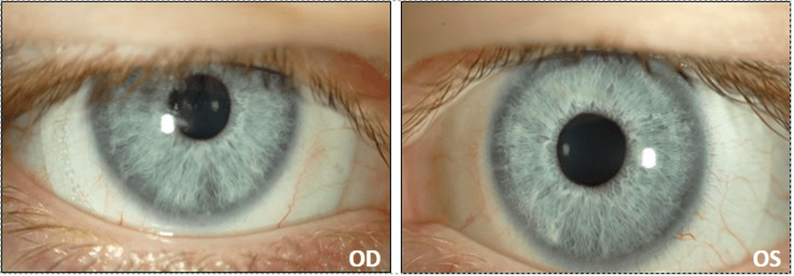
The patient’s visual complaints were consistent with that of residual HOAs. To quantify the degree of HOAs with her presenting PDs, corneal aberrometry scans were obtained for each eye. Aberrometry scans confirmed residual HOAs, left eye worse than right (Figure 1).
Figure 1: iTrace aberrometry scans demonstrating residual HOAs with patient’s presenting PROSE device for the right (OD) and left (OS) eye. Residual HOAs were more pronounced for the left than right eye, consistent with patient’s chief complaint.
After further discussion of clinical findings, the patient confirmed symptoms of suction with lens wear including aching and soreness as well as rebound redness after lens removal for both eyes. The decision was made to modify the fit for both eyes, with the goals of improving comfort by mitigating suction and optimizing vision by attempting to correct for the residual HOAs.
Refitting/ PROSE Retreatment
With PROSE retreatment, the diameter was increased by 0.5mm for both eyes to provide a wider haptic edge to better distribute the weight of the device for each eye (Figure 2). This was done to reduce the amount of suction with prolonged lens wear. Each lens was also converted from a quadrant-specific haptic design to an eight-meridian haptic design for optimal customization and fitting. With retreatment, the patient’s rebound hyperemia and complaints of discomfort after PD removal were resolved.
Figure 2: Finalized PROSE fit for the right (OD) and left eye (OS) in 19.0mm designs.
HOA Correction with PROSE
Once the fit was stable for each eye, we pursued HOA correction. Aberrometry scans over the patient’s optimized PDs were acquired under dilated conditions to better quantify the degree of HOAs in scotopic conditions.
The aberrometry data was incorporated into the PROSE design for each eye. A repeat refraction was performed over the wave-front guided PROSE devices, which was included in the final design. The patient reported significant improvement in daily visual function, with improvement of “ghosting” in the left eye and complete resolution of ghosting for the right eye.
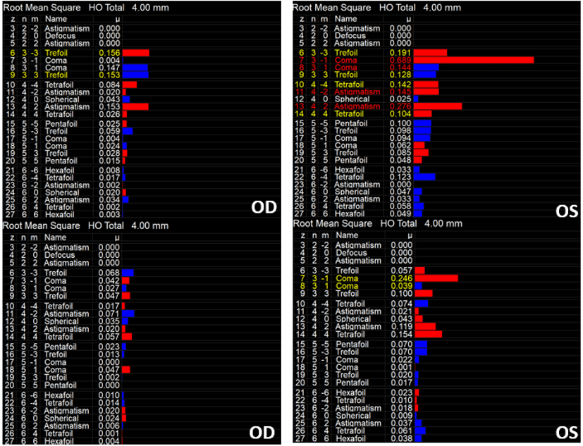
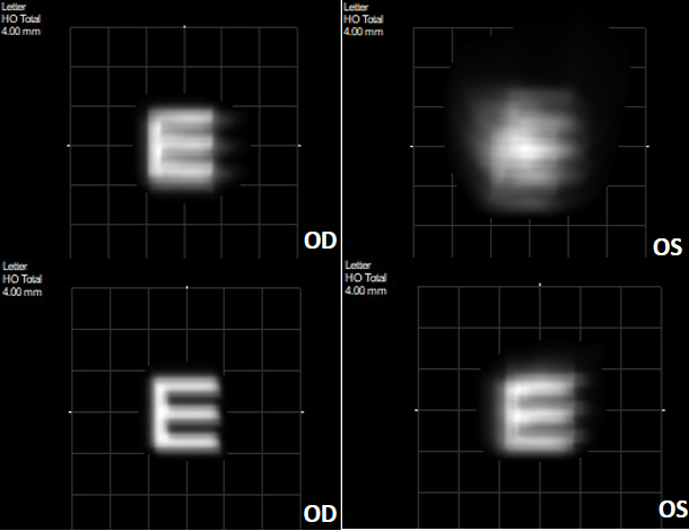
Repeat aberrometry scans were obtained over the final wavefront corrected PDs which confirmed significant reduction in HOAs. The most notable improvement was observed for coma, which can be the most visually significant in KCN patients (Figures 3 and 4).
Figure 3: iTrace aberrometry scans with patient’s presenting PROSE devices (top) for the right (OD) and left (OS) eye, compared to patient’s finalized PROSE devices (bottom) for the right (OD) and left (OS) eye.
Figure 4: iTrace visual function analyzer (VFA) scan demonstrating the vision quality with the patient’s presenting right (OD) and left (OS) PD (top) versus the patient’s finalized wavefront-guided devices (bottom). There is a notable improvement in quality of vision based on the VFA for each eye.
Discussion
Higher Order Aberrations and Scleral Lenses
Bringing wavefront-guided scleral lenses involves various considerations, including determining who may be an appropriate candidate. The HOA profile of each patient is as unique as their fingerprint – no two eyes are the same. The recipe for HOA correction with scleral lenses has four key ingredients:
Patient must have some vision improvement potential.
Improved vision with pinhole.
Symptoms of HOAs which can be quantified as HOAs based on aberrometry scans.
Access to an aberrometer, with reliable aberrometry scans.
A rotationally and tangentially stable scleral lens design.
Much like the need for a toric soft CL to be stable to function properly, a wavefront-guided scleral lens also needs to be stable while on the eye.
Access to a wavefront-guided lens design with an appropriate manufacturing lab.
Conclusion
Although specialty CLs including scleral lenses can greatly improve visual function and correct for some degrees of HOAs, residual HOAs can haunt patients’ visual function as they experience symptoms like blur, distortion, doubling, shadowing, glare, and ghosting. Correcting for HOAs can further improve visual function in patients suffering from KCN; therein, potentially further reducing the need to pursue invasive corneal surgery such as penetrating keratoplasty.
As technological advances continue to unfold and HOA correction through wavefront-guided technology becomes increasingly available to practitioners, the future is looking increasingly bright for patients.
1. Schornack MM. Scleral lenses: A literature review. Eye Contact Lens. Published online 2015. doi:10.1097/ICL.0000000000000083 2. Sabesan R, Johns L, Tomashevskaya O, Jacobs DS, Rosenthal P, Yoon G. Wavefront-guided scleral lens prosthetic device for keratoconus. Optom Vis Sci. 2013;90(4):314-323. doi:10.1097/OPX.0b013e318288d19c 3. Chalita MR, Xu M, Krueger RR. Correlation of Aberrations with Visual Symptoms Using Wavefront Analysis in Eyes after Laser in situ Keratomileusis. J Refract Surg. 2003;19(6):682-686. doi:10.3928/1081-597X-20031101-13 4. Naderan M, Shoar S, Kamaleddin MA, Rajabi MT, Naderan M, Khodadadi M. Keratoconus Clinical Findings According to Different Classifications. Cornea. Published online 2015. doi:10.1097/ICO.0000000000000537 5. Marsack JD, Ravikumar A, Nguyen C, et al. Wavefront-guided scleral lens correction in keratoconus. Optom Vis Sci. Published online 2014. doi:10.1097/OPX.0000000000000275 6. Koppen C, Kreps EO, Anthonissen L, Van Hoey M, Dhubhghaill SN, Vermeulen L. Scleral Lenses Reduce the Need for Corneal Transplants in Severe Keratoconus. Am J Ophthalmol. Published online 2018. doi:10.1016/j.ajo.2017.10.022 7. Chahal JS, Heur M, Chiu GB. Prosthetic Replacement of the Ocular Surface Ecosystem Scleral Lens Therapy for Exposure Keratopathy. Eye Contact Lens. 2017;43(4):240-244. doi:10.1097/ICL.0000000000000265
Thank you to Bita Asghari for contributing to Global Insight.
[us_separator size=”small” show_line=”1″]
[us_image image=”9240″]
[us_separator size=”huge”]
Dr. Asghari is an optometrist and the Associate Director of Clinical Education for BostonSight. She also serves as adjunct clinical faculty for MCPHS school of Optometry and New England College of Optometry (NECO). She completed her Doctorate in Optometry from Southern California College of Optometry, graduating summa cum laude. Prior to joining BostonSight, Dr. Asghari completed her PROSE clinical fellowship, as well as her post-doctoral residency in Cornea & Contact Lens and Ocular Disease from The Ohio State University Havener Eye Institute.