Tag: keratoconus

The Changing Landscape of Keratoconus, the Irregular Cornea & Contact Lens Correction

[us_post_title][us_image align=”center” size=”us_600_0″ image=”10509″][us_post_date]

By Eef van der Worp

Keratoconus, without exception, has always been our number one indication for specialty lenses, particularly scleral lenses, for both pre and post-surgical intervention. Studies from the University of Utrecht, Netherlands, by Godefrooij et al.¹ show that the incidence of keratoconus may be much more common than originally thought. The annual incidence of keratoconus is reported to be 1 in 7,500 for the relevant age category or 13.3 cases per 100,000. The prevalence of keratoconus in the general population was estimated to be 1:375. These values appear to be 5-fold to 10-fold higher than previously reported. Two reasons for this may be the ever-increasing use of corneal topographers in practices globally and increased awareness of the condition. 

So, because keratoconus is here to stay – as is secondary ectasia (often resulting from refractive surgery) – this will remain a key consideration in every specialty lens practice. For severe irregularities in stage 3 and stage 4 keratoconus, scleral lenses come to the rescue – as they bridge over the entire cornea, resting on the scleral portion (the conjunctiva, really) on the ocular surface. This explains, at least in part, the vast success of scleral lenses over the last decade.

Cross-Linking

However, the landscape of the irregular cornea in keratoconus seems to be changing. First came the development of corneal cross-linking (CXL), which has now been around in Europe for 20 years (human studies of UV-induced CXL began in 2003 in Dresden, Germany). CXL does not cure or diminish keratoconus but can help stop further progression at a given stage. The primary purpose and literal definition of CXL is to halt the progression of ectasia, although this is not always entirely feasible. Typically, the earlier in life that the procedure is performed, the better the result. That said, the best candidates for CXL are individuals with a progressive ectatic disease of the cornea, so ECPs must monitor that first before the procedure is considered. In other words, the cornea is already irregular to at least some degree by then, and preventative CXL is not feasible.

So, while across Europe, and increasingly in other parts of the world, CXL is becoming the standard of care for young patients with progressive ectatic disease, it is most likely we will see more keratoconic patients. However, it is to be expected that we will see corneal changes in the earlier stages of the condition. This may impact what correction methods we apply: glasses and soft (specialty) lenses may become more common as the first-line method of vision correction.

Lamellar Corneal Transplants

There have been major developments in corneal transplants for keratoconus and other diseases. Not too long ago, a penetrating keratoplasty – replacing the entire cornea in its full thickness with donor tissue – was the standard procedure. Now, an array of lamellar techniques in which only a portion of the cornea is replaced by donor tissue is increasingly used. This can provide many advantages, one being that the corneal irregularities are far less prominent, as a part of the patients’ original cornea remains intact. 

In the late nineties, a technique of deep anterior lamellar keratoplasty (DALK) was developed by the Netherlands Institute for Innovative Ocular Surgery (NIIOS)². The thought behind this technique is to replace only the diseased part – that is, the connective tissue layer of the cornea – and to fasten it with sutures. The healthy part of the cornea is ‘left in place,’ avoiding many of the disadvantages of a full-thickness corneal transplantation. 

Bowman Layer Transplant 

Recently the NIIOS developed another new surgical technique: Bowman layer transplantation. Just as with CXL, Bowman layer transplantation intends to prevent further deformation of the cornea and delay the need for a corneal transplant. This technique is indicated when the cornea is too thin for CXL. With a Bowman layer transplantation, a thin layer of donor cornea (limited to just a donor Bowman layer) is implanted between the connective tissue layers of the cornea, making the original cornea become firmer. Sometimes even two donor Bowman layers are placed on top of each other on the receiving eye for that purpose. 

Soft Lenses to the Rescue

All these techniques may lead to a significant reduction of severe irregular corneas. This may also mean that scleral lenses will not always be necessary. Eye care practitioners around the globe increasingly consider a soft lens as an excellent first option after glasses fail to provide adequate visual acuity. The advantages of this are obvious: scleral lenses are large and handling is a known issue, wearing time is not always optimal, and limitations such as mid-day fogging, redness and potential intraocular pressure increase are eliminated. Especially the ease of use, comfort and cost are regularly mentioned as a plus for soft lenses. 

This doesn’t always work with standard (off-the-shelf) soft lenses. First, the overall sagittal height of the ocular surface regularly goes beyond 4,000 microns over a 15mm chord in these corneas. Because of this, standard commercially available lenses on the extreme right-hand side of the Pacific SAG charts are recommended³. If this is not feasible, ‘out-of-standard’ soft lenses with higher SAG values can be considered. These have virtually no limit in SAG value height. Customized soft lenses based on corneal topography – with tangential peripheral shapes, almost like a scleral soft lens – are on the market as well (also with monthly replacement in silicone hydrogel materials). The question is: Does it still correct or at least mask the corneal irregularities enough for acceptable vision?

Masking

If the answer to the latter question is negative, there is a newer category of soft specialty lenses specifically for keratoconus. These lenses generally have increased central thicknesses, typically in the 300-450 micron range (a standard soft disposable lens may be 80 microns thick) to (partially) mask the irregularity of the cornea to achieve acceptable vision. Although these lenses can be made in silicone hydrogel materials, the Dk/t is lower because of the ‘t’ (thickness) of the lens, and patients wearing these lenses should be monitored for hypoxia. Some lenses can be ordered in 100-micron steps to increase or decrease thickness. Some lenses also have the option for ‘sector management,’ in which one quadrant can be ordered flatter or steeper (like quadrant-specific corneal or scleral lenses) to align the lens better with the keratoconic eye. 

Illustrative of the latest surge in interest in soft specialty lenses for the irregular cornea is that recently some new soft keratoconus lens designs have entered the market. Some of these also try to improve vision further by using aspheric front optics. Trial sets in some of these lenses are also marked in sagittal height increments. 

In Summary 

So, while on the one hand, corneas don’t get nearly as irregular as they used to for a variety of reasons, on the other hand, the lens material, design and optics of soft specialty lenses are improving. The landscape of the keratoconic/irregular cornea, therefore, seems to be changing. And with that, the landscape for specialty lenses in that indication is also transforming, with a potentially more prominent place for soft lenses in that arena. It may be time to adhere to that – in clinical practice and from an industry perspective. 

Educational Series & the Future

The educational grant over 2022, kindly supported by Contamac, has allowed the author of this article to provide generic specialty lens education around the globe for a better understanding of what we are doing with different specialty lens modalities and to potentially take advantage of all of the possibilities available to us. Topics over the past year included mostly scleral lenses, the use of corneal topography, scleral topography, and myopia management with specialty lenses. But increasingly, soft lens fitting in all its aspects (how to better fit standard lenses, when to go to out-of-standard lenses and soft lenses for the irregular cornea) is gaining more and more attention. According to the annual Eurolens survey published in Contact Lens Spectrum (Jan 2023), at least 86% of all lenses fitted today are soft. But we can’t fit all these patients with standard lenses. The future looks bright for specialty lenses, especially when it comes to soft specialty lenses. We are just beginning to scratch the (ocular) surface. Stay tuned. 

[us_separator show_line=”1″ size=”small”]
Godefrooij DA, de Wit GA, Uiterwaal CS, Imhof SM, Wisse RP. Age-specific Incidence and Prevalence of Keratoconus: A Nationwide Registration Study. Am J Ophthalmol. 2017 Mar;175:169-172
Netherlands Institute for Innovative Ocular Surgery: https://www.niios.com/melles-cornea-clinic/treatments/
van der Worp E, Lampa M, Kinoshita B, Fujimoto MJ, Coldrick BJ, Caroline P. Variation in sag values in daily disposable, reusable and toric soft contact lenses. Cont Lens Anterior Eye. 2021 Dec;44(6):101386. doi: 10.1016/j.clae.2020.11.006. Epub 2021 Jan 8. PMID: 33423977.
[us_separator show_line=”1″ size=”small”]
[us_image image=”9904″ size=”us_600_600_crop” has_ratio=”1″ ratio=”3×4″]

Eef van der Worp is an educator and researcher. He received his optometry degree from the Hogeschool van Utrecht in the Netherlands (NL) and has served as a head of the contact lens department at the school for over eight years. Eef received his PhD from the University of Maastricht (NL) in 2008. He now runs his own research & education consultancy ‘Eye-Contact-Lens’ which is based in Amsterdam (NL). He is a fellow of the AAO, BCLA and the SLS, a lifetime fellow of IACLE and an honorary life member of ANVC. He is on the education committee for the GSLS and the OVN.  Eef is adjunct assistant Professor at Pacific University College of Optometry (Oregon, USA), and adjunct Professor at the University of Montreal University College of Optometry (CA) and he is lecturing extensively worldwide and is a guest lecturer at a number of Universities in the US and Europe.

[us_separator size=”large”]
[us_separator size=”small” show_line=”1″]
[us_post_navigation in_same_term=”1″]
[us_sharing providers=”facebook,twitter,linkedin” align=”right”]
Tags: , , , , ,

Refitting Success: Reducing Suction and Maximizing Oxygen Permeability

[us_post_title][us_post_date][us_separator size=”small”]

By Karen Carrasquillo, OD, PhD, FAAO, FSLS, FBCLA

Case Summary

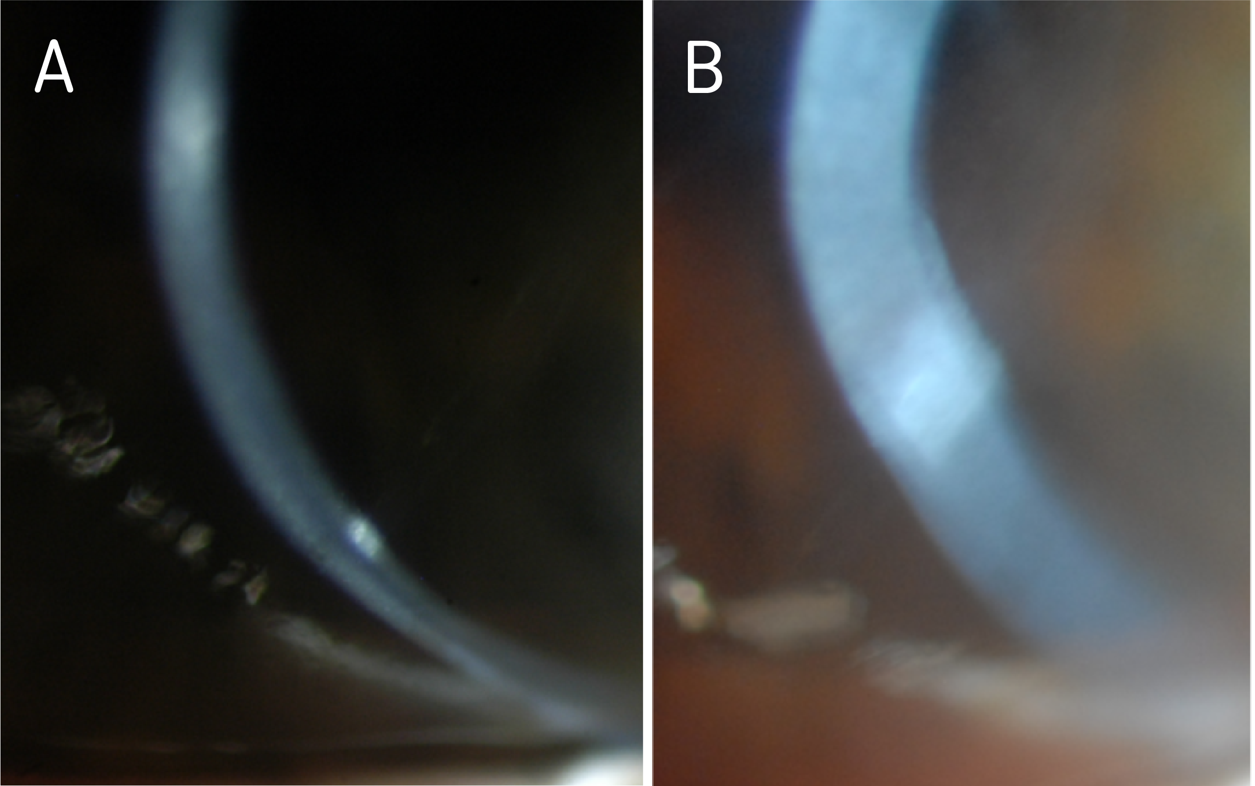
Case of a 60-year-old woman with a history of keratoconus and dry eye syndrome in both eyes, history of corneal scraping in the left eye 25 years prior, history of post hydrops left eye, and questionable potential history of amblyopia left eye. She had a history of successful PROSE treatment since 2010 (11 years of PROSE device wear) but underwent penetrating keratoplasty (PKP) in the left eye in December 2019; secondary to diffuse opacification (Figure 1) and limited best corrected visual acuity.

Prior to PKP in the left eye, she had been wearing 18.5mm, quadrant-specific PROSE devices in both eyes, with added front-surface astigmatism in the right eye and with resultant best corrected visual acuity (BCVA) of 20/30, PH 20/25-2 and 20/100, PH 20/60–, right eye and left eye respectively. With prescribed polycarbonate spectacles to correct residual astigmatism in the left eye and to serve as protection (especially for the better-seeing eye), BCVA in the left eye could be improved to 20/70. The patient wore the spectacles as needed, depending on daily activities and especially during night driving.

Reducing Suction and Maximizing Oxygen Permeability

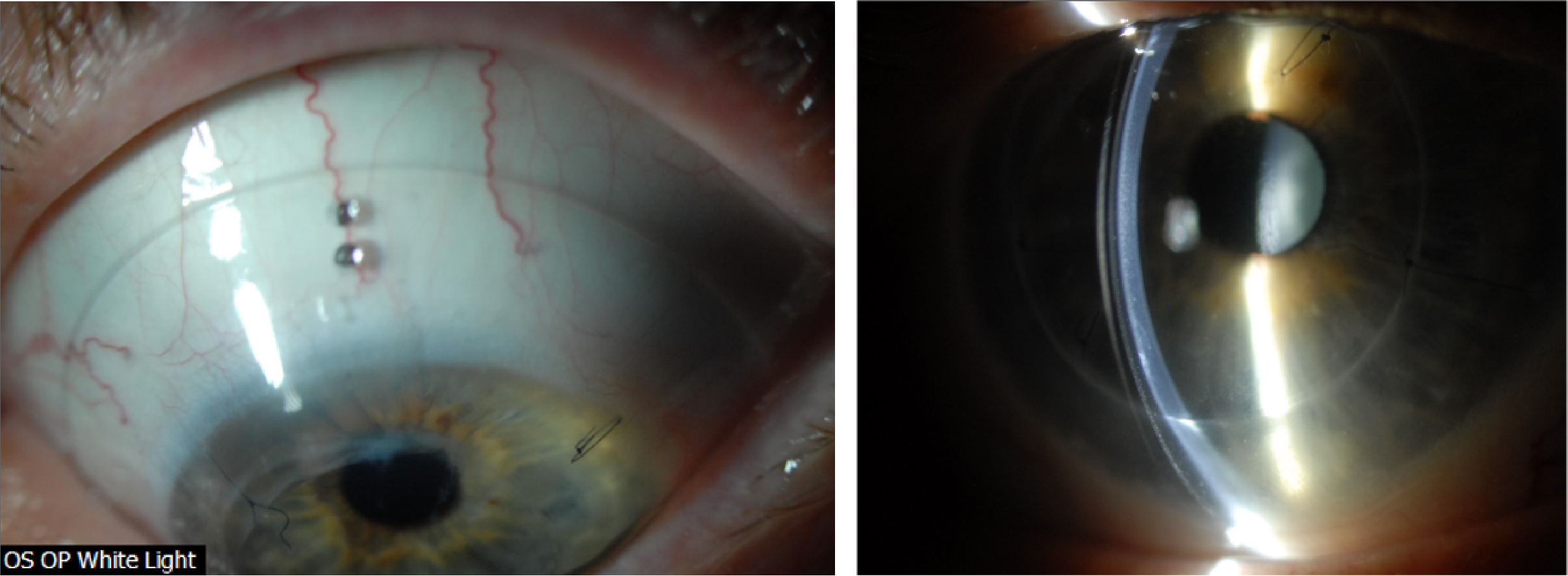
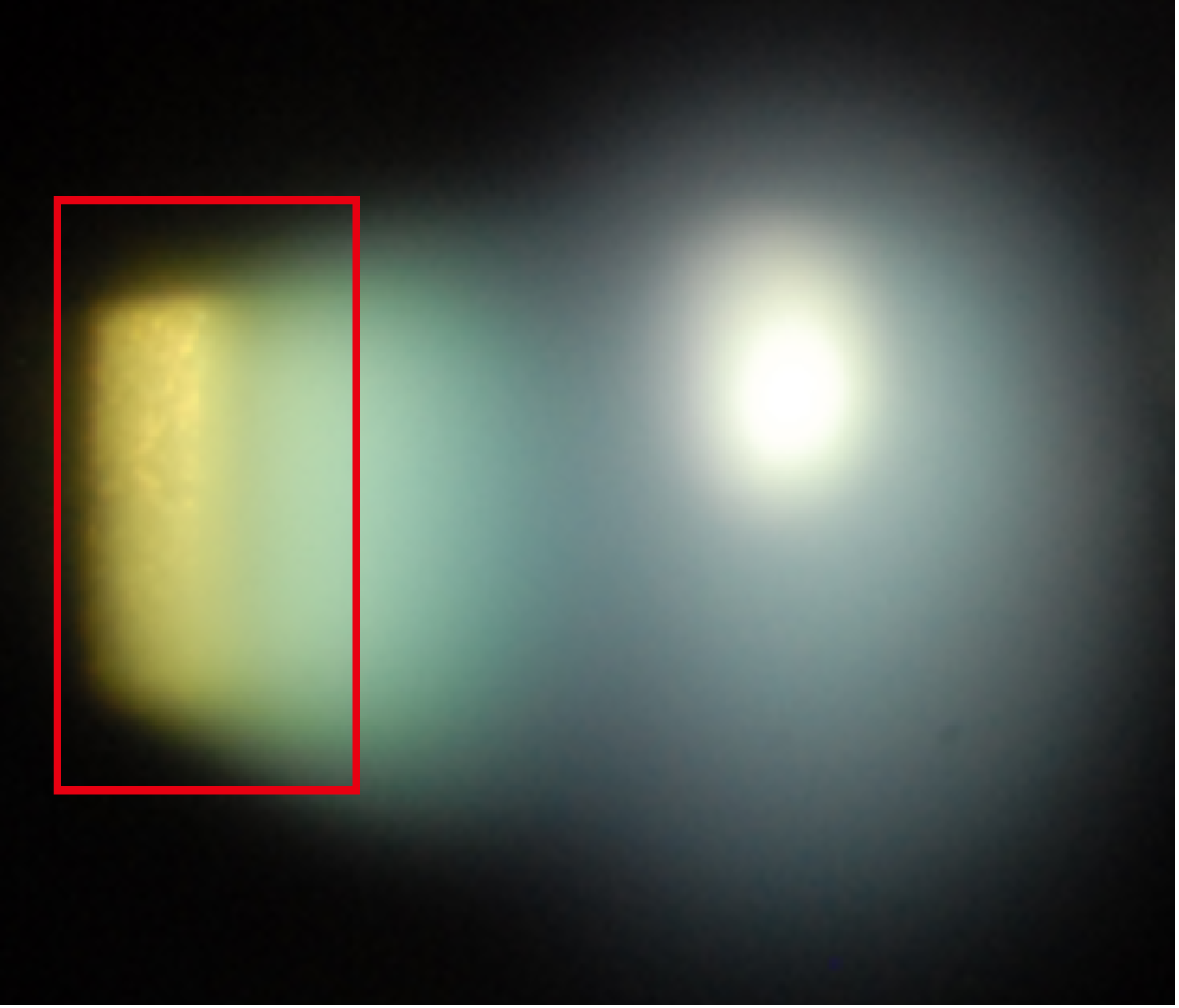
Figure 1. Diffuse haze and opacification in the left eye, as seen on optic section (A) and wide beam (B), prior to PKP.

After corneal transplantation and having been cleared to resume scleral device wear in the left eye, she was referred to our clinic for refitting.

Evaluation After Penetrating Keratoplasty

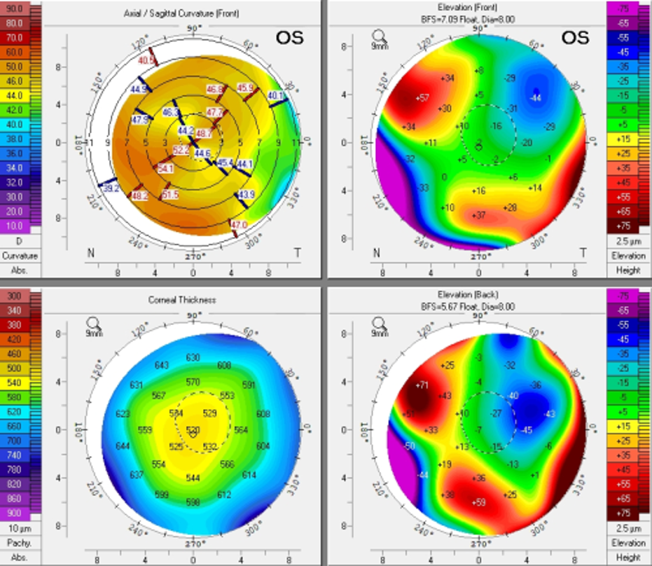
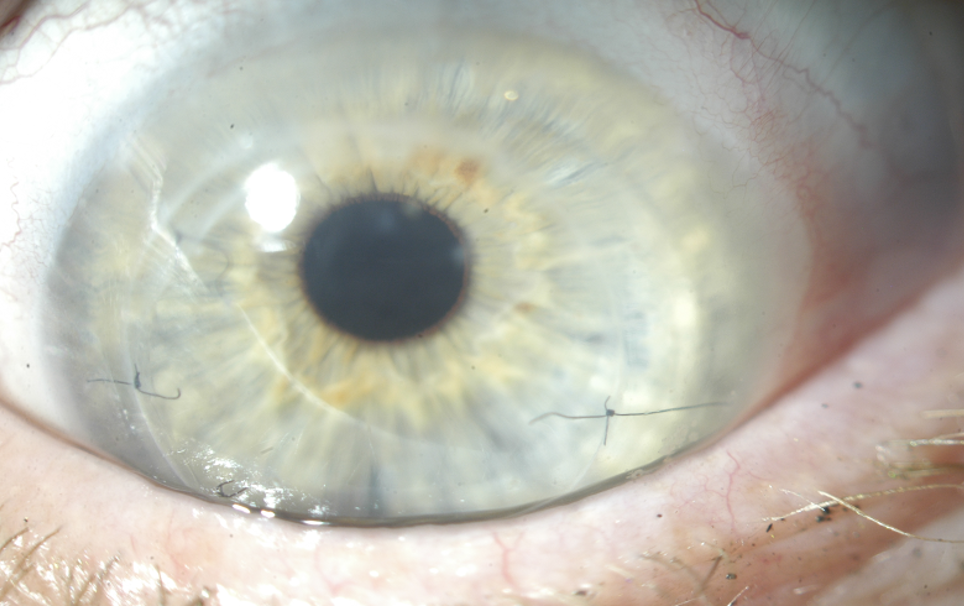
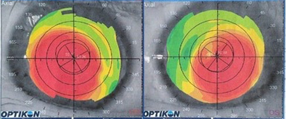
Initial evaluation after PKP OS (Topography shown in Figure 2A), showed a clear and compact graft, with interrupted sutures (Figure 2B). There was no evidence of subepithelial infiltrates, microcystic edema, no keratic precipitates, or stromal diffusion after corneal staining. The patient was on Prednisolone acetate 1% gtts QD and as sutures were removed, she was prescribed Moxifloxacin 0.5% TID in the left eye. Evaluation was also remarkable for cataracts in both eyes, not visually significant at the moment. Best corrected vision during this visit in the right eye was 20/40, PH 20/30-2 and uncorrected visual acuity in the left eye was CF at 3’, PH 20/200.

Figure 2A. Pentacam images s/p PKP in the left eye.

Figure 2B. Presentation of left cornea, status post corneal graft during baseline evaluation for refitting.

Refitting

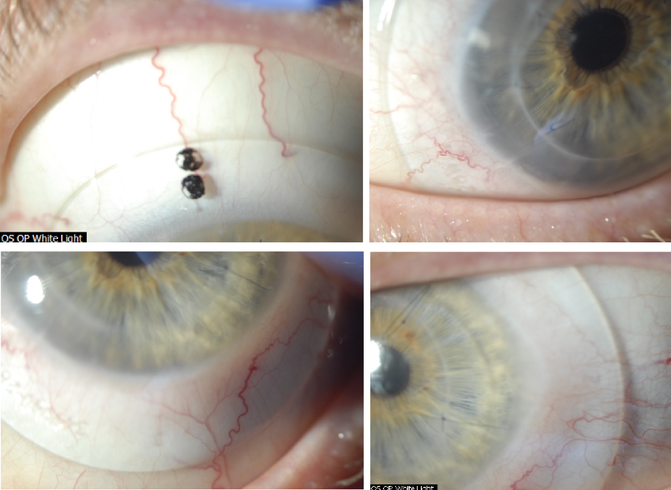
We started a new fit, deciding to keep the patient in the habitual diameter of 18.5mm. Because the cornea was now grafted, we opted to design the lens using Optimum Infinite material (180 Dk) so that together with a center thickness of 0.25mm, oxygen permeability was maximized. Best documented corrected visual acuity in the left eye was 20/30-2 with the new 18.5mm device. While the patient was elated about the newfound visual potential, she reported sensation of pressure during device wear in the left eye – despite no objective evidence of significant compression at the haptics (Figure 3).

Figure 3. Initial fitting endpoint with an 18.5mm PROSE device after PKP OS.

An attempt was made to flatten the haptics, modify the area along the mid-haptic portion to minimize mid-haptic compression and obtain better alignment. Despite these efforts, the patient continued to report pressure sensation with device wear and difficulty with device removal at the end of the day. These symptoms are traditionally associated with lens compression and suction with increased wear time. After reviewing the findings and the thought process with the patient, the decision was made to increase the lens diameter to 19.5mm to provide a larger surface area on which the device could rest. Figure 4 shows the new fitting endpoint with the 19.5mm, 8-meridian quadrant-specific device. One can observe good haptic alignment and the eye is white and quiet, with a nice and even distribution of lens clearance over the grafted cornea.

Reducing Suction and Maximizing Oxygen Permeability

Figure 4. Fitting endpoint with an 8-meridian, quadrant-specific 19.5mm PROSE device after PKP OS.

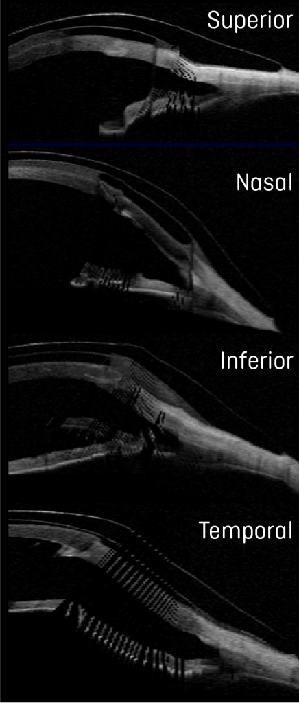
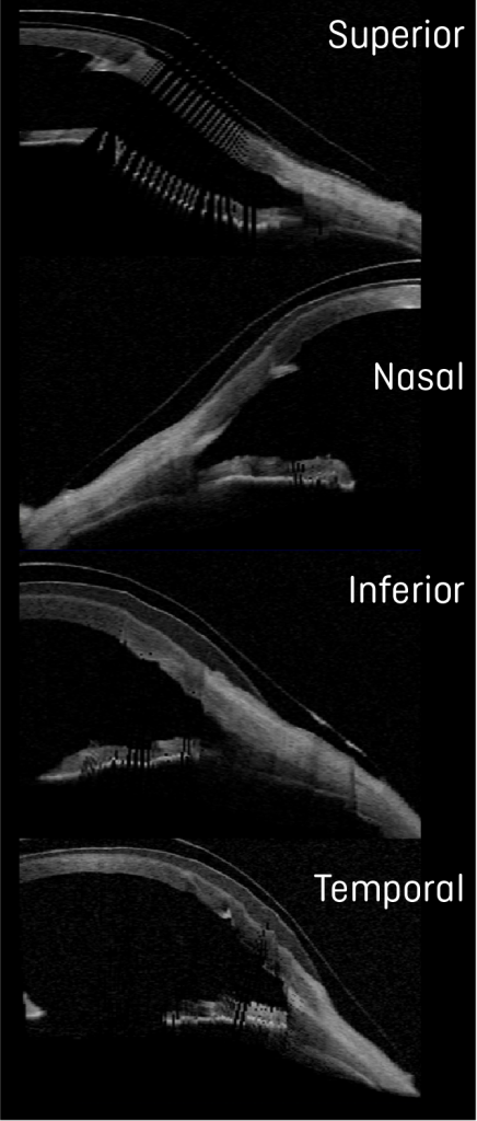
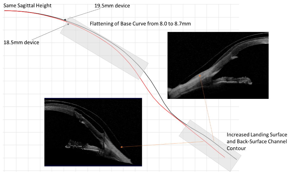
Comparison OCTs show the increased landing surface area when going from an 18.5 (Figure 5) to a 19.5mm device (Figure 6). This attempt to maximize weight distribution over a larger surface area has proven efficacious in our clinical practice when dealing with compression symptoms and device intolerance. While maintaining the same sagittal height, we additionally flattened the base curve to accommodate the more oblate nature of the cornea after transplant as shown in Figure 7.

Reducing Suction and Maximizing Oxygen Permeability

Figure 5. Ocular Coherence Tomography (OCT) of an 18.5mm device in the different orthogonal meridians of the left eye.

Reducing Suction and Maximizing Oxygen Permeability

Figure 6. Ocular Coherence Tomography (OCT) of a 19.5mm device in the different orthogonal meridians of the left eye.

Figure 7. Graphical images of 18.5 and 19.5mm, juxtaposed to show the changes to the lens design based on parameter changes using spline curve technology and as observed by OCT. The nasal meridians corresponding to the flattest meridian in the eye are shown for comparison.

Interestingly, while we were optimizing the fit and after reporting improvement in comfort with increased lens size, the vision in the left eye dropped to the 20/50-20/60 range over the course of a couple of months. During all evaluations, we carefully monitored for signs of microcystic epithelial corneal edema, signs of rejection, etc. and at no point were evidence of these found. Given the history of previous cataracts and the fact the patient was on chronic low dose steroids, we performed an undilated assessment of the human lens in the left eye. Photo documentation revealed the development of a posterior subcapsular cataract (PSC) (Figure 8), which was later confirmed to be visually significant during dilated assessment by her corneal specialist. The patient decided to move forward with cataract extraction with hopes to improve vision.

Figure 8. Evidence of PSC formation, during undilated assessment.

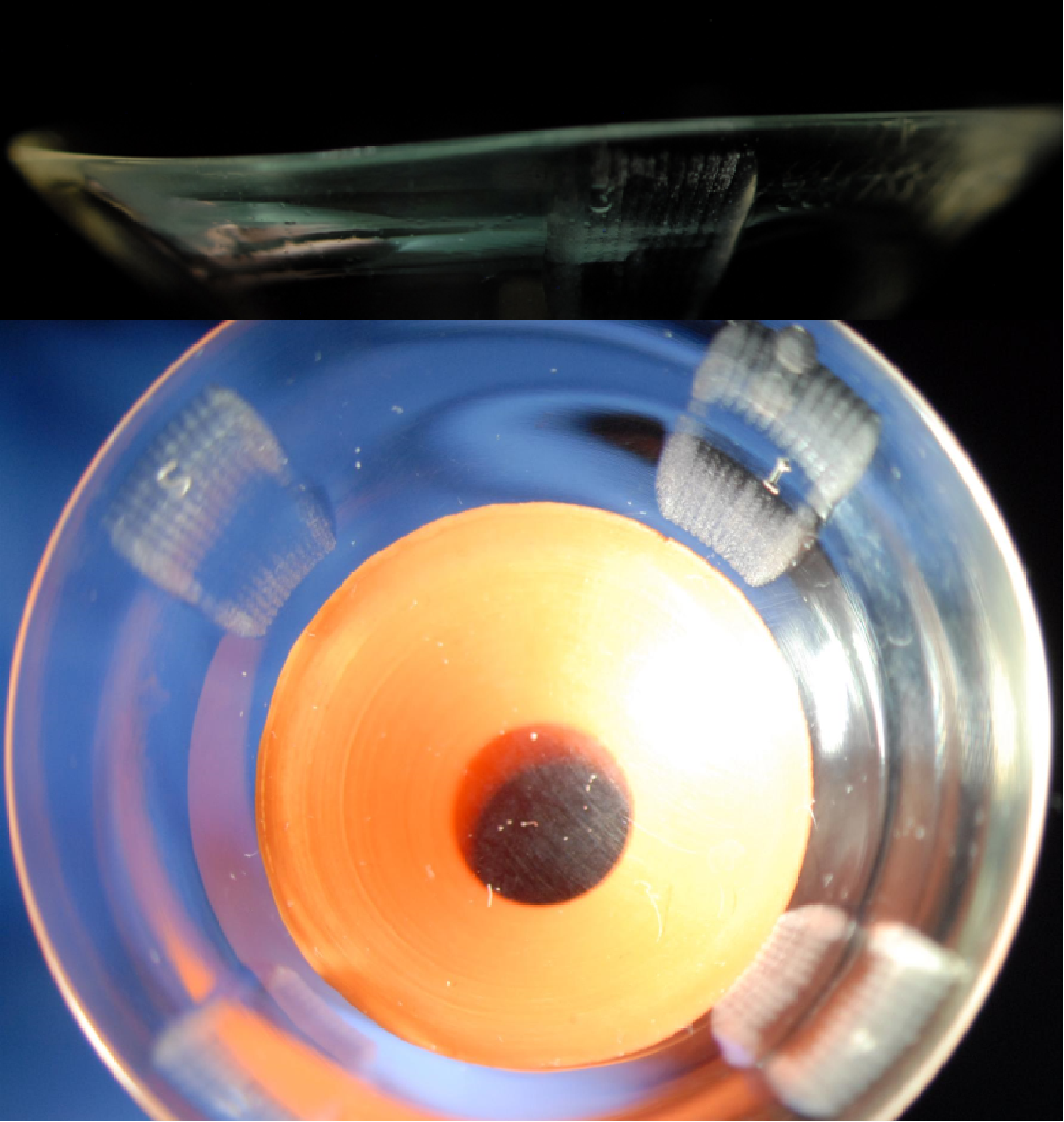
Before cataract surgery, we decided to challenge the current 19.5mm design hoping to obtain a comfortable and physiologically compatible lens design. After the patient wore the lens for one week, she reported that the previous symptoms of pressure and difficulty with lens removal had improved, but they were still present. To further minimize compression and suction symptoms, we added 4 back-surface haptic channels to soften further the landing of the haptic surface from bearing harshly on the conjunctiva and prevent a seal off effect (Figure 9).

Reducing Suction and Maximizing Oxygen Permeability

Figure 9. A 19.5mm, 8-meridian quadrant-specific PROSE device with the addition of 4 back-surface haptic channels to minimize suction and improve comfort.

Discussion and Final Outcomes

Long-term outcomes and benefits of scleral lenses after penetrating keratoplasty have been reported in the literature – while most patients benefit from improvement in visual acuity, complications with scleral lenses, i.e. microcystic corneal edema can ensue in this patient population.1 Special consideration should be taken when fitting or refitting grafted corneas with scleral lenses to minimize the risk of corneal edema, erosions and ultimate rejection. Strategies to promote fluid-ventilation like avoiding tight-fitting edges, lens seal-off and adding back-surface haptic channels can be especially important.1-5 Clinical practice has also shown that the addition of back-surface channels can have the potential to minimize lens suction over time and lead to improvement in physiological endpoints.5-7

In this case, with an increase in scleral lens landing surface area, customized haptic in an 8-meridian quadrant-specific manner and the addition of 4 back-surface haptic channels, not only are we promoting fluid-ventilation to optimize physiological endpoints, but we have a reported significant improvement in comfort. The choice to manufacture lenses in Optimum Infinite material potentiates the oxygen permeability to the grafted cornea. With the final lens after cataract surgery, the patient was able to achieve 20/25+2 best corrected visual acuity with an average comfortable daily wear of up to 12 hours.

By undertaking strategies to progressively soften the landing over the conjunctival surface to reduce or eliminate lens compression over time and minimize suction we were able to achieve a successful refitting outcome.


1. Severinksi, B. et. al. (2014) Scleral contact lenses for visual rehabilitation after penetrating keratoplasty: long-term outcomes. Cont Lens Ant Eye, 37, 196-202.
2. Rosenthal, P. Croteau, A (2005) Fluid-ventilated, gas-permeable scleral contact lens is an effective option for managing severe ocular surface disease and many corneal disorders that would otherwise require penetrating keratoplasty. Eye Cont Lens, 31(3), 130-134.
3. Severinksy, B et. al. (2010) Current applications and efficacy of scleral contact lenses – a retrospective study. J Optom, 3(3), 158-163.
4. Stason WB, Razavi M, Jacobs DS, et al. (2010) Clinical benefits of the Boston ocular surface prosthesis. Am J Ophthalmol, 149, 54–61.
5. Carrasquillo, KG, Byrnes, S (2018) Corneal edema and scleral lenses. Cont Lens Spect, 33, 34-41.
6. Barnett, M (2018) Mind the suction. Scleral Lens Monthly, Nov 2018.
7. Carrasquillo, KG (2020) Mind the suction II. Scleral Lens Monthly, Jul 2020.

Thank you to Dr Karen G.Carrasquillo for contributing to Global Insight.

[us_separator show_line=”1″ size=”small”]
[us_image image=”9526″]

Dr. Karen G. Carrasquillo is Vice President of Clinical and Professional Affairs at BostonSight. In addition, she is an adjunct clinical professor at the New England College of Optometry, an adjunct clinical professor at the school of optometry, MCPHS University, Advisory Board member for the Gas Permeable Lens Institute (GPLI), Fellow of the American Academy of Optometry (FAAO), Fellow of the Educational Society of Scleral Lenses (FSLS), and Fellow of the British Contact Lens Association (FBCLA). She is the founder and Program Chair of FitAcademy, an educational retreat for Cornea and Contact Lens Residents, and is also a planning committee member for the International Congress of Scleral Contacts (ICSC) Meeting and Global Ophthalmic Women (GLOW) meeting.

[us_separator show_line=”1″ size=”small”]
[us_post_navigation in_same_term=”1″]
[us_sharing providers=”facebook,twitter,linkedin” align=”right”]
Tags: , , , , ,

Backtracking From Scleral Lenses: When Custom Soft Lenses Get the Job Done

[us_post_title][us_post_date]

by Priya Patel, OD.

Introduction

Scleral lenses provide significant visual and therapeutic advantages for patients suffering from a variety of corneal conditions: inclusive of kerato-ectasias, penetrating keratoplasties, and varying severities of corneal irregularities.

This advancement in the specialty lens realm has expanded to the use of sclerals, yet previous technologies are still capable of providing comparable comfort and vision. 

Custom soft lenses have potential to significantly improve vision without the need for complex designs, and small diameter gas permeable lenses can achieve similar visual outcomes as sclerals at a fraction of the cost. Custom soft lenses can be entirely customizable from base curve, diameter, power, to even center thickness to improve upon masking corneal irregularities. Often in clinical practice, there exist simpler solutions to complex problems.

Case Report

A 46-year-old White male teacher with a history of keratoconus OD>OS presents to our office seeking scleral lenses. Patient JB has a history of gas permeable (GP) lens intolerance and has successfully avoided PKP (penetrating keratoplasties) in both eyes. The continuous irritation in his GP lenses and the fear they can pop out at any moment, has him seeking out scleral lenses.

His corneal shape shows most significant steepening directly over the visual axis, which would be well vaulted by a prolate design. Utilizing the Zenlens (Alden) Prolate fitting set, JB was fit into a 16.0 diameter scleral lens in both eyes, achieving BCVA 20/25 OD/OS. The initial fitting period was completed without any significant complications, and JB was happy to have clear, comfortable vision. At the one year mark after his initial scleral lens fitting, JB started to develop significant redness after 4 hours of lens wear with concurrent fogging, which only resolved after reinsertion of his lenses. After lens removal, the chronic irritation persisted. This led to intolerance of his lenses.

Zenlens Prolate
Scleral Lens
Right Eye Left Eye
SAG/Base Curve/Diameter (μ/mm/mm) 4950/6.70/16.0 4950/6.70/16.0
Power -4.75-2.00×150 -6.25-1.25×070
Limbal Clearance Curve (LCC) +60 STD
Advanced Peripheral System H = 7 steps flat
V = STD
2 steps flat
Visual Acuity 20/25 20/25

JB became more frustrated, and he pleaded for any lens option, which would yield a similar visual outcome, but improve his comfort. We were sceptical we would be able to achieve this, but in an effort to exhaust all options available,  we switched him into a custom soft lens modality in both eyes.  To optimize comfort and reduce the chance of redness, fogging, irritation after lens removal as JB previously experienced, he is fit into the NovaKone Toric OD and Biofinity XR Toric OS. The custom soft lens selection was based on the poor BCVA (20/80+) attainable through refraction: the customizable center thickness of the Novakone lens would allow greater control in neutralizing irregular astigmatism in the right eye. The left eye was correctable to an adequate level (20/25+) with glasses so a monthly disposable lens had potential to correct JB to a level at which he would be functional. 

Custom Soft Lenses

K Values:

OD: 55.15@121/53.26@031

OS: 50.88@053/49.52@143

Custom Soft Lenses

OD: Novakone Toric lens, rotated 15 degrees temporally (markings are highlighted with yellow dashes).
OS: Biofinity XR Toric lens, showing no rotation (lens marking is highlighted with a yellow dash).

 
Refraction  
OD -12.75-1.75×025      20/80+
OS -0.50-4.00×145       20/25+
Custom Soft Lens Right Eye Left Eye
Name Novakone Toric Biofinity Toric XR
Base Curve/
Diameter (mm)
7.8/8.4 IT=2 8.7/14.5
Power -7.00-0.75×020 -2.00-2.75×160
Visual Acuity 20/20- 20/20-2

These changes in lens modality improved his contact lens wearing experience and significantly simplified his daily lens wear routine. He has been in his new lenses for over a year with no degeneration in vision, and no recurrences of discomfort or irritation.

Discussion

Scleral lenses are a mainstay as a rehabilitative and therapeutic contact lens option, but alternatives exist for patients from mild to advanced cases of corneal irregularities. Taking into account comfort, lens handling capabilities, alternative specialty lens considerations can lead to improved outcomes as demonstrated in this case.

Scleral lenses require hand steadiness, ability for full control of fine motor skills, and neck mobility. Patients suffering from tremors, neck pain, or mobility issues often have trouble inserting scleral lenses on their own, but have minimal issues with soft and GP lenses. Improper insertion of scleral lenses can lead to air bubbles, and discomfort with lens wear. On the contrary,  minimal risk is associated with difficulties with soft lens insertion .¹

Scleral lenses are associated with an increased risk of hypoxia if the tear film layer between the corneal surface and the back surface of the lens exceeds 200nm.²?³ Soft and GP lens lenses pose a lower risk for creating a hypoxic environment even for fragile corneas.

Utilizing higher modulus soft lenses allow some masking of irregular astigmatism for better visual outcomes. Although in this case, since JB achieved adequate vision through a refraction  in his left eye, we were able to fit him in a lower modulus standard soft disposable lens. If he experienced difficulties with the quality of his vision in the left eye, a higher modulus monthly disposable lens would be his next option. His right eye, however, warranted a lens with a greater center thickness to mask his irregular astigmatism. Custom soft lenses are entirely customizable, inclusive of increasing the central thickness to enhance the masking of any irregularities.⁴

Scleral lenses will often be a superior lens for patients suffering from contact lens discomfort due to lens intolerance (without underlying dry eye disease). As they remain steady on the ocular surface, the optics can be decentered to allow for improved correction of higher order aberrations versus a soft lens or a GP lens.⁶

Conclusion

Soft contact lenses and gas permeable lenses are viable options for therapeutic lens correction. Scleral lenses can be considered an excellent choice as a troubleshooting lens, but it cannot be the only option available for patients who are seeking a specialty contact lens solution. When presented with a patient who is not comfortable in their scleral lenses, often a creative solution is warranted to find an alternative solution.


1. Fadel D, Toabe M. Scleral Lens Issues and Complications Related to Handling, Care and Compliance. JCLRS [Internet]. 2018Oct.26 [cited 2021Jan.13];2(2):e1-e13. Available from: http://www.jclrs.org/index.php/JCLRS/article/view/24
2. Compañ V, Oliveira C, Aguilella-Arzo M, Mollá S, Peixoto-de-Matos SC, González-Méijome JM. Oxygen diffusion and edema with modern scleral rigid gas permeable contact lenses. Invest Ophthalmol Vis Sci. 2014 Sep 4;55(10):6421-9. doi: 10.1167/iovs.14-14038. PMID: 25190661.
3. Michaud L, Vincent S. SCLERAL LENSES AND HYPOXIA: A BALANCED APPROACH. https://www.clspectrum.com/issues/2019/october-2019/scleral-lenses-and-hypoxia-a-balanced-approach. Published 2019.
4. Şengör T, Aydın Kurna S. Update on Contact Lens Treatment of Keratoconus. Turk J Ophthalmol. 2020;50(4):234-244. doi:10.4274/tjo.galenos.2020.70481.
5. Downie, L. E., and Lindsay, R. G. (2015), Contact lens management of keratoconus. Clin Exp Optom, 98, 299– 311. doi: 10.1111/cxo.12300.
6. Waleed Ali Abou Samra, Amani E. Badawi, Hanem Kishk, Ayman Abd El ghafar, Mohamed M. Elwan, Hossam Youssef Abouelkheir, “Fitting Tips and Visual Rehabilitation of Irregular Cornea with a New Design of Corneoscleral Contact Lens: Objective and Subjective Evaluation”, Journal of Ophthalmology, vol. 2018, Article ID 3923170, 8 pages, 2018. https://doi.org/10.1155/2018/3923170

Thank you to Dr Priya Patel for contributing to Global Insight.

[us_separator size=”small” show_line=”1″]
[us_image image=”9323″]
[us_separator]

Dr. Priya Patel received her Doctorate of Optometry from SUNY. She joined the premier practice in NYC and Long Island: Farkas, Kassalow, Resnick and Associates upon graduation, where she focuses clinically on speciality contact lens fittings. Dr. Patel routinely helps patients with keratoconus and other corneal disorders, as well as managing progressive nearsightedness with contact lenses (myopia management).  She also designs and prescribes custom contact lenses such as scleral lenses, hybrid lenses, and multifocal lenses. Dr. Patel has authored articles in the area of specialty contact lenses, and hosted workshops on practice management related to specialty lenses. She is currently residing in London.

[us_separator show_line=”1″ size=”small”]
[us_post_navigation in_same_term=”1″]
[us_sharing providers=”facebook,twitter,linkedin” align=”right”]
Tags: ,

Bilateral Keratoconus: An Interesting Use of an EyeBrid AirKone Hybrid Lens with Tangible Hydra-PEG Coating

[us_post_title][us_post_date]

by Dimitri Guimond, MSc in Optical Sciences and Optometry

The patient is a 25-year-old male supermarket worker and a keen sportsman (football). He consults his ophthalmologist every two years at his city-centre location. He has reduced visual acuity that cannot be improved with glasses. He has no particular medical-surgical history. He was referred to a University Hospital Centre (CHU) specialising in the management of keratoconus. After multiple examinations, he was referred by his specialist physician to a contact lens fitting centre for a first-time contact lens fit.

Clinical Presentation

During the eye test, the best-corrected visual acuity (BCVA) with glasses was measured at 20/200 OD and 20/20- OS.

Examination of the anterior segments on the slit lamp revealed:

·  A tear film with few inclusions, regular tear flow

·  Grade 1.5 bulbar and palpebral conjunctival injection on the Efron scale

·  Grade 1.5 eyelid roughness on the Efron scale

·  Corneal thinning more pronounced on the apex OD with the presence of Vogt’s striae.

·   OS presents some thinning and no Vogt’s striae visible with the slit lamp.

The corneal topography below highlights the different stages of the evolution of the keratoconus, its morphology and its location.

Bilateral Keratoconus

Typical appearance of a small, very pointed keratoconus with a strong peripheral flattening indicates a form of keratoconus that can be related to the Nipple Cone (NC) family.
Keratometry: 6.34/097*5.83 e=1.66

Bilateral Keratoconus

Here we see a typical form of this keratoconus. This side is less severe, with gentler flattening.
Keratometry: 7.30/067*7.12 e=0.60


Lens Fitting

Given the patient’s active lifestyle, we chose hybrid lenses in order to optimise the centration and stability of the lenses.

In order to meet the needs of this patient, we opted for a contact lens geometry with a design particularly suited to these two different stages of keratoconus, the AirKone geometry.

Comfort was improved through the addition of the Tangible Hydra-PEG treatment.

To give maximum flexibility in fitting options, the laboratory recommends carrying out the initial fitting assessment with a corneal GP lens design, noting the parameters, and then ordering that lens with the addition of the hybrid skirt. This means that the practice does not need to maintain duplicate fitting sets for GP keratoconus and hybrid keratoconus lenses, for example.

The initial fit was done with an AirKone corneal GP lens to assess the correct alignment using fluorescein. Following the fitting rules, r0=average K, we tried the following fitting set lenses:

Keratoconus Hybrid Lens

AirKone: 6.10/8.70/-13.00 EL+1.0
Central fluorescein shows the lens is too flat by 0.1mm
Optimal peripheral fluorescein
Overcorrection: -1.25 20/20-

Keratoconus Hybrid Lens

AirKone: 7.20/9.00/-3.00 EL 0.0
Central fluorescein shows the lens is aligned over the cone
Optimal peripheral fluorescein
Overcorrection: -2.75 20/20+

Based on the GP results, we followed the guidelines provided by the laboratory to order the EyeBrid AirKone lenses. In order to ensure good surface wettability, we specified the addition of the Tangible Hydra-PEG surface coating when ordering the lens.

Keratoconus Hybrid Lens

OD: EyeBrid AirKone 6.00 / 14.90 / -15.00. Edge lift +1.0 J(skirt) 0.0

Keratoconus Hybrid Lens

OS: EyeBrid AirKone 7.20 / 14.90 / -5.75. Edge lift 0.0 J(skirt) 0.0

The pictures show a good fit. BCVA rose to 20/20- OD and 20/20 OS. Surface wetting is good, and the patient reports good comfort. He wears his lenses for his professional and leisure activities for about 14 hours a day. He cares for his lenses with a hydrogen peroxide product and puts 2 drops of a hyaluronic acid-based comfort solution into each lens before putting them in his eyes. He puts his lenses in with a handling ring and removes them with a suction cup.

Patient Follow Up

After 3 weeks of wearing, the examination performed with the lenses on eye showed good stability, and slit lamp observations were unremarkable. We checked the integrity of the patient’s corneal epithelium after removing the Eyebrid lens: the corneas show no epithelial complications and no corneal staining.

Keratoconus Hybrid Lens

Conclusion

Thanks to its excellent centration, stability, visual quality and comfort, the EyeBrid hybrid lens offers real solutions for both fitters and patients, regardless of the corneal morphology. Combined with a Tangible Hydra-PEG surface treatment, the lenses wet extremely well and are deposit-resistant while limiting friction with the eyelids. All these elements play in favour of improving patient comfort and allow the patient to continue wearing contact lenses.

Discussion

Although this patient was new to contact lenses, the same fitting philosophy can be applied to many existing, happy GP wearers using many different GP lens designs, who are getting older, whose tear film is changing, and who are experiencing reduced comfort levels. By keeping the same lens design, but simply adding a skirt, the patient can experience improved comfort levels.

Note: Eyebrid lenses have a GP centre manufactured from Optimum Extra (Dk 100), surrounded by a silicone hydrogel skirt manufactured in Definitive 50.

Thank you to Dimitri Guimond for contributing to Global Insight.

[us_separator size=”small” show_line=”1″]
[us_image image=”9344″]
[us_separator]

Dimitri is an optometrist and the director of Professional Services at LCS laboratory, contact lens manufacturer of hybrid, scleral, RGP, Ortho-K and soft lenses.
He has also taught at the University of Paris Orsay in optometry, and actively participates in the development of continuing education in contact lenses for ophthalmologists, orthoptists, optometrists and opticians. He regularly consults in private practice as well as in university hospital centres.

[us_separator show_line=”1″ size=”small”]
[us_post_navigation in_same_term=”1″]
[us_sharing providers=”facebook,twitter,linkedin” align=”right”]
Tags: ,