Successful Visual Restoration of Presbyopia with a New Customized Multifocal Corneo-Scleral Contact Lens
by David P Piñero, PhD
Introduction
The use of scleral contact lenses has grown significantly in the last year, with an exponential increase in research on this issue.1 Although there is a consensus about most of the aspects of contact lens management among practitioners with more than 5 years of scleral lens experience,2 there are some aspects that require more research, such as the management of multifocality for presbyopia correction. Recently, a new model of multifocal corneo-scleral contact lens has been commercially released by Laboratorios Lenticon S.A. (Madrid, Spain), Presbycustom, providing the option of customizing not only the fitting parameters to adjust the lens over the cornea and sclera, but also the type of multifocality induced. We report the results obtained with this new modality of multifocal scleral contact lens in a specific case not tolerating multifocal soft contact lenses due to poor and fluctuating visual quality.
Case report
A 45-year old man attended the Contact Lens Unit of the Department of Ophthalmology of the Vithas Medimar International Hospital (Alicante, Spain) asking for a contact lens solution for his presbyopia. He was a contact lens user for 16 years. He noticed some difficulties at near due to the development of presbyopia and tried to find a contact lens solution for the compensation of this condition. However, the results obtained with different models of soft multifocal contact lenses (5 contact lens models were previously tried) have been unsatisfactory as the visual quality with them was not good, with fluctuation of vision with most of them. On the first examination, the following clinical data were obtained:
| Right Eye | Left Eye | |
| Subjective Refraction | +1.75 -0.50 x 70 | +1.00 |
| Corrected Distance Visual Acuity (CDVA) | 0.00 logMAR | 0.00 logMAR |
| Near Addition | +1.00 | +1.00 |
| Corrected Near Visual Acuity (CNBA) | 0.00 logMAR | 0.00 logMAR |
| Biomicroscopy | No alterations detected | No alterations detected |
| Scotopic Pupil Size | 6.0 mm | 6.0 mm |
| Mean Keratometry | 41.17 D | 41.2 D |
| Ocular Spherical Aberration (6 mm pupil) | 0.26 µm | 0.21 µm |
No significant corneal shape alterations were found on corneal topography, with values of corneal asphericity and high order aberrations within a normal range (Figure 1). Considering the peculiarity of the case, we decided to fit a customized multifocal scleral contact lens. Specifically, the Presbycustom contact lens was fitted, which is a corneo-scleral contact lens made of highly gas permeable material (Roflufocon D, Optimum Extra, 100 Dk). This lens has a customized design according to the anterior corneal geometry, the level of ocular high order aberrations and the peculiarities of pupil dynamics. The corneal area has a variable diameter with a fixed back surface asphericity and a central anterior surface asphericity which is modified according to the aberrometric induction required. Specifically, the increase in the depth of focus achieved with the contact lens is generated by customizing the induction of primary and secondary spherical aberration.3,4
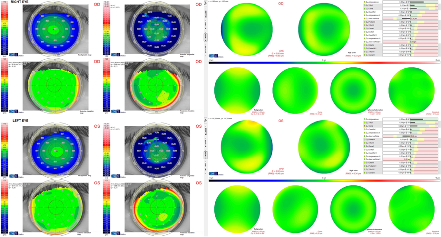
Figure 1. Corneal topographic analysis with the Sirius system (CSO, Firenze) in both eyes of the case report described.
Different contact lenses were tried until obtaining the best fit with the following lenses in right and left eyes, respectively:
| Right Eye | Left Eye | |
| Lens Diameter | 13.5 mm | 13.5 mm |
| Corneal Radius | 7.65 mm | 7.46 mm |
| Scleral Radius | 7.55 mm | 7.40 mm |
| Optical Power | -1.00 D | -3.00 D |
| Anterior Eccentricity | 0.63 | 0.55 |
| Posterior Eccentricity | 0.63 | 0.63 |
With these lenses, the fluorogram was correct, showing a central tear pooling, with a paracentral alignment and a significant edge clearance (Figure 2). Likewise, a smooth peripheral bearing of the lens was observed by means of optical coherence tomography (Figure 3).
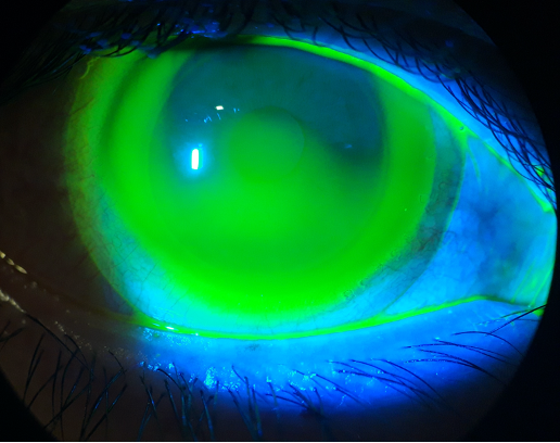
Figure 2. Fluorogram obtained in the right eye of the case fitted.
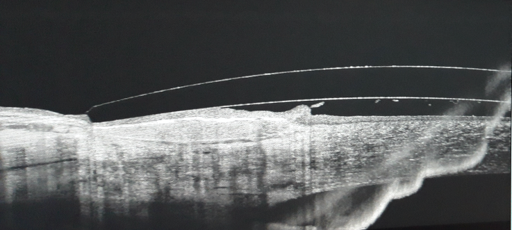
Figure 3. Peripheral bearing of the lens observed by means of optical coherence tomography.
At 1 month of contact lens wear, the visual acuities with the contact lens were 0.00 logMAR and 0.10 logMAR at distance and near (40 cm), respectively, in both eyes (overrefraction: plano in both eyes). Figure 4 shows the binocular defocus curve measured, confirming the significantly enlarged depth of focus achieved.
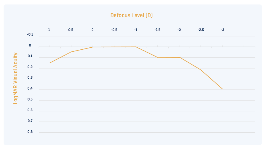
Figure 4. Binocular defocus curve measured in the patient.
The patient has been successfully wearing the contact lens for 2 years, with maintenance of the results and good levels of tolerance.
Conclusion
The use of customized scleral contact lenses allows the practitioner to optimize the presbyopia correction in any type of presbyopic patient. The selection of an appropriate combination of primary and secondary spherical aberration of opposite signs according to the aberrometric status of the eye is an effective tool to optimize the depth of focus required for presbyopia correction in each individual case. This preliminary result should be corroborated in the long term as well as in a larger sample size.
1. Davies I. Scleral publications & contact lens category growth. Cont Lens Anterior Eye 2019; 42: 234-235.
2. Van der Worp E, Bornman D, Ferreira DL, Faria-Ribeiro M, Garcia-Porta N, González-Meijome JM. Modern scleral contact lenses: A review. Cont Lens Anterior Eye 2014; 37: 240-50.
3. Yi F, Iskander R, Collins M. Depth of focus and visual acuity with primary and secondary spherical aberration. Vis Res 2011; 51: 1648-1658.
4. Benard Y, López-Gil N, Legras R. Optimizing the subjective depth-of-focus with combinations of fourth- and sixth-order spherical aberration. Vis Res 2011; 51: 2471-2477.
Thank you to David P Piñero, PhD for contributing to Global Insight.

David P Piñero has a degree in Optics and Optometry from the University of Alicante (1998) and a degree in Information and Documentation from the Open University of Catalonia (2006). He obtained his PhD Degree at the University of Alicante in 2010 with the doctoral thesis entitled “Characterization and modelling of the effect of the intrastromal ring segments de in ectatic corneas”. Likewise, he obtained the qualification of University Specialist in Pre- and Post-surgical Optometry by the University of Valladolid in 2001. Currently, he is professor/researcher at the Department of Optics, Pharmacology and Anatomy of the University of Alicante, associate editor of Journal of Optometry, Journal of Ophthalmology and BMC Ophthalmology, and optometrist at the Department of Ophthalmology of the Vithas Medimar International Hospital (Alicante, Spain). He has published more than 300 papers in peer-reviewed journals, most of them on the cornea, and participated in 20 book chapters. He has also participated in more than 20 research projects.